FINITE ELEMENT ANALYSIS
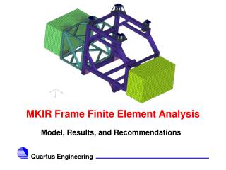
Development of a Biomimetic Finite Element Model to Study the Intervertebral Disc Diseases and Regeneration. Author * ANDRÉ CASTRO Supervisors: Luís Alves, Paulo Flores, António Completo * apgcastro@dem.uminho.pt. PhD grant SFRH/BD/63882/2009. INTRODUCTION

FINITE ELEMENT ANALYSIS
E N D
Presentation Transcript
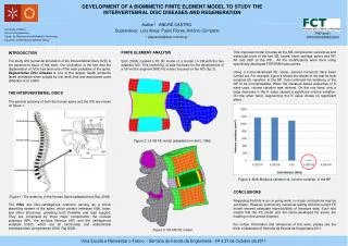
Development of a Biomimetic Finite Element Model to Study the Intervertebral Disc Diseases and Regeneration Author* ANDRÉ CASTRO Supervisors: Luís Alves, Paulo Flores, António Completo * apgcastro@dem.uminho.pt PhD grant SFRH/BD/63882/2009 INTRODUCTION The study and numerical simulation of the Intervertebral Discs (IVD) is the paramount issue of this work. Our motivation is the fact that the degeneration of IVDs has been one of the main problems of the spine. Degenerative Disc Disease is one of the largest health problems faced worldwide when judged by lost work time and associated costs (Shankar et al. 2009). THE INTERVERTEBRAL DISCS The general anatomy of both the human spine and the IVDare shown on figure 1. Figure 1: The anatomy of the Human Spine (adapted from Raj, 2008) The IVDs are fibro-cartilaginous cushions serving as a shock absorbing system of the spine, which protect vertebrae (VB), brain, and other structures, providing both flexibility and load support. They are composed by three major components: the nucleus pulposus (NP), the annulus fibrosus (AF) and the cartilaginous endplate (CEP), which are all functionally and anatomically interdependent (Jongeneelen 2006, Raj 2008). FINITE ELEMENT ANALYSIS Smit(1996) created a FE 3D model of a human L4 VB with the two adjacent IVD. This model (fig. 2) was the basis for the development of a full motion segment (MS) FE model, focused on the IVD (fig. 3). Figure 2: L4 VB FE model (adapted from Smit, 1996) Figure 3: 3D MS FE model This improved model includes all the MS components: cancellous and trabecular bone of the two VB, facets, facet cartilage layers and NP, AF and CEP of the IVD . All the modifications were done using specifically developed FORTRAN subroutines. Using a home-developed FE solver, several numerical have been carried out. For example, figure 4 shows the results of the test for bulk modulus (K) variation, in the NP. One confirmed the tendency of the NP to be incompressible. When the literature based properties of K were used, volume variation was minimal. On the one hand, only a large decrease in the K value caused a significant volume variation. On the other hand, augmenting the K value shows no significant effect. Figure 4: Bulk Modulus variation vs. volume variation, in the NP CONCLUSIONS Regarding that this is an on-going work, no major conclusions may be yet drawn. However, preliminary numerical testing with the current FE model showed adequate reproducibility of literature data. Such fact means that the FE model and the home-developed FE solver are heading on the aimed direction. For further information and references of this work, please see the book of abstracts of Semana da Escola de Engenharia 2011.