Day 2
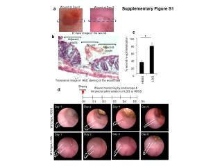
d. Wound at Day 0. Wound at Day 2. a. Supplementary Figure S1. c. En face image of the wound. b. *. 100 80 60 40 20 0. Adjacent crypts. Wound. Adjacent crypts. % wound re-epithelialization. *. Transverse image of H&E staining of the wound bed. HBSS LGG. Biopsy Injury.

Day 2
E N D
Presentation Transcript
d Wound at Day 0 Wound at Day 2 a Supplementary Figure S1 c En face image of the wound b * 100 80 60 40 20 0 Adjacent crypts Wound Adjacent crypts % wound re-epithelialization * Transverseimage of H&E staining of the wound bed HBSS LGG Biopsy Injury Wound monitoring by endoscope & Intrarectal adminstration of LGG or HBSS d D0 D1 D2 D3 D4 D5 D6 Day 1 Day 2 Day 4 Day 6 Wild type, HBSS Day 4 Day 6 Day 2 Day 1 Wild type, LGG
Supplementary Figure S2 a Media 50 500 5,000 LGG (2.5107 cfu/ml) LGG (2.5107cfu/ml) fMLF (500nM) fMLF: (nM) 5106 1.25108 2.5107 Media LGG : (cfu/ml) To-pro 3 (DNA) ROS (CM-H2DCF-DA) ROS (CM-H2DCF-DA) 15’ 30’ 60’ 5’ b wound wound wound wound HBSS wound wound wound wound LGG
Supplementary Figure S2 (continued) c 2.5 x 10⁸ cfu 2.5 x 10⁹ cfu 2.5 x 10¹⁰ cfu Control 2.5 x 10⁷ cfu wound wound wound wound wound wound WT wound wound wound wound wound FPR1 KO 50 nM 500 nM 5 uM 50 uM Control d wound wound wound wound wound WT wound wound wound wound wound FPR1 KO
WT, LGG Supplementary Figure S3 a Min 0 10 15 20 P-FAK 125 kDa P-paxillin 68 kDa Actin 45 kDa b Adjacent crypts Adjacent crypts Staining for EdU chase assay to detect cellular migration Day 0 1 2 4 Biopsy wound & labeling of cells by EdU Wound Wound bed Fpr1-/- Wild type c HBSS, 1hr D * * HBSS 6hr % migration LGG 6hr WT HBSS WT LGG Fpr1 -/- HBSS Fpr1 -/- LGG EdU (chase 6 hrs) DNA
Supplementary Figure S4 LGG a 0 10 15 20 Min P-ERK 44/42 kDa Actin 45 kDa b Actin P-ERK WT, NAC + LGG Fpr1-/-, LGG Nox1-/-IEC, LGG WT, LGG WT, HBSS 15 min 60 min
a Supplementary Figure S5 I.P. injection of EdU 2hr prior to wound bed collection & EdU staining to detect proliferation Biopsy injury Day 0 Day 1 Day2 Day4 B EdU DNA Wild Type, Day 1 Wild Type, Day 2 Wild Type, Day 4 Adjacent crypts Adjacent crypts Adjacent crypts Wound Wound Wound b Wild Type, LGG, Day 2 Wild Type, HBSS, Day 2 Adjacent crypts Adjacent crypts Adjacent crypts Adjacent crypts Wound Wound EdU DNA c Fpr1-/-, LGG Nox1-/-IEC , LGG Wild type, HBSS Wild type, LGG Wound Wound Wound Wound DNA Ki67 (+)