Assessing Risk from Medical Radiation
920 likes | 1.16k Vues
Assessing Risk from Medical Radiation. Elizabeth H. Ey, M.D. Medical Director Radiology Dayton Children’s Medical Center. Content of presentation and pictures courtesy of Thomas Slovis, M.D. and The Society for Pediatric Radiology. Survey.

Assessing Risk from Medical Radiation
E N D
Presentation Transcript
Assessing Risk from Medical Radiation Elizabeth H. Ey, M.D. Medical Director Radiology Dayton Children’s Medical Center
Content of presentation and pictures courtesy of Thomas Slovis, M.D. and The Society for Pediatric Radiology
Survey Has anyone here read a news report of radiation exposure from medical imaging?
Survey Has anyone had a patient or family member ask them about radiation exposure from medical imaging?
Survey Has anyone had a test question in medical education (or CME) regarding radiation exposure to patients from medical imaging tests?
Questions • What is our natural background level of radiation? • What is the radiation dose of a 1 view chest radiograph? • What is the radiation dose of a 1 view abdomen radiograph? • What is the radiation dose of a CT scan? • Head CT dose? • Abdomen CT dose?
Answers • Natural occurring background radiation 1 mrad/day • Chest, one view, child, skin dose 3-15 mrad • Abdomen, one view, child, skin dose 50 mrad • CT Head, child, minimal dose, CTDI 2000 mrad • CT Abd, child, CTDI >1000 mrad
What is the increased risk of death from cancer from 1 CT scan performed in a child? • A The increased risk of cancer death from a CT scan is 0. • B The increased risk of cancer death is between 1 in 1000 to 1 in 5000 over a lifetime. • C The increased risk of cancer death is 1 in 1 million over a lifetime.
Answer • B It is estimated that an abdominal CT scan results in 1/1000 to 1/5000 excess risk of cancer at a later date.
What is the current lifetime risk of • developing cancer (US)? • dying of cancer (US)?
Lifetime risk of cancer (US) Developing Dying Male 44.05% 23.24% Female 37.6% 19.65%
Medical Radiation in Medicine • When indicated it can diagnose illness • Noninvasive, painless, fast, extremely accurate • But like any medication or therapy • Too much radiation can lead to deleterious effects • DETERMINISTIC effect –linear, direct, ex: skin reddening • Any radiation dose can cause lethal effect – cancer • STOCHASTIC effect – non-linear, random, takes time to see
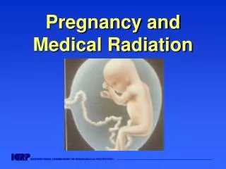
Think of radiation as a medicine • Effects are lifelong and cumulative • Particularly severe effect in infants and children • Especially when adult doses are used in children • Age dependent – younger patient more severely effected • No dose of radiation can be considered completely safe • Linear non-threshold effect
ALARA Concept for Pediatric Radiation Dose • As • Low • As • Reasonably • Achievable
Medical Radiation in Children • History of Radiology • Basic dosimetry • Biology of radiation effects • Unique issues with radiation in children • Use of appropriate techniques • Joint efforts with healthcare providers
History • Dec 28, 1895: Roentgen submits manuscript describing his discovery of “x-ray” to the Physical Medical Society of Wurzburg: manuscript printed and distributed in 3 days • Jan 9, 1896: manuscript appears in Vienna Press • Jan 23, 1896: manuscript appears in Nature, in England • Jan 23, 1896: Roentgen presented paper to Physical Medical Society of Wurzburg • By mid 1896, fluoroscopy was in widespread practice • From clinical bench to widespread use in 6 months
Wurzburg Medical-Physics Society 1896 Dr. Kohler, famous anatomist, having hand X-rayed by Roentgen at the meeting.
Roentgen received the first Nobel Prize in 1901 for his discovery
Application of Radiation Sciences for Medical Diagnosis: • Tremendous benefits • Risks became evident with increasing use
Side Effects of Radiation in Humans Started being reported within months of discovery of x-ray
Public Spectacle: Side Effects • Deep sunburn • Hair loss • Bloodshot eyes, vision impaired • Transient effects
Practitioners in X-ray techniques were the first to show the long term effects of radiation exposure
Monument to Martyrs in X-ray and Radium Physics – 1936 Hamburg • Albers Schonberg • Madame Curie • Caldwell • Codman
Br J Radiol 2001; 74: 507- 519 Berrington A, Darby SC, Weiss HA, Doll R • Research on 100 years of data on health of radiologists in Great Britain • 1897-1954 – 41% excess of cancer deaths in practitioners of radiology • 1954-1997 – zero excess mortality from cancer in the practitioners of radiology
Learned biologic effects of radiation • Applied what we learned to protect ourselves • It worked • But have we done enough? • Have we protected our patients enough?
2. Basic dosimetry • Dose units • Measures of dose • Conversions
Methods of Measuring Radiation Dose • Widely varied and difficult to compare • Entrance skin dose • Exit dose • Dose area product (DAP) • Organ dose –specific to radiosensitive organ • Radiation output measured within a phantom • CT dose index (CTDI) • Dose equivalent • Effective dose
Radiation Dose Measurements Used for Risk Assessment • Absorbed Dose – Gray or Gy (previous rad) • Risk assessment for a specific organ or tissue • Difficult to measure and not very useful • Effective dose equivalent – Sievert or Sv (previous rem) • Non-uniform exposure to organ or region • Expression of risk equivalent to whole body exposure • CT scanner dose units not useful • CTDI vol and DLP determined by phantom • Not helpful for assigning risk without conversion
CTDI – CT Dose Index • Reported on scanner consoles • Based on phantom (16 or 32 cm diameter) • Only represents the dose to the phantom based on CT parameters selected • Does not indicate dose to the child in the CT scanner • Conversions of CTDI to effective dose are only rough estimations for children • e.g. no age based chest modifications
Dose Chart • 1 Gy = 100 rads = 1 Sv • 10 mGy = 1 rad = 10 mSv • 0.01 mGy = 1 mrad = 0.01 mSv
Effective Dose • It is a radiation dose quantity • It is a computation based on: Organ dose and radiosensitivity Weighting factors • It is not a risk number Huda, W Pediatric Radiology 2002: 32; 272-279
Types of Biological Effects from Radiation • Deterministic effects • Stochastic effects
Deterministic Effect • Seen with high radiation dose • Severity of effect is dose dependent: • There is a threshold below which dose the effect is NOT seen. • Examples: skin burns, hair loss
AJR July 2001 - Skin burns from cardiac interventional procedures
Stochastic Effect • Low dose, random effect • Non dose dependent: • Risk of the effect is dose dependent but the severity of the effect is not. • Example: Risk of cancer increases with increasing dose but the severity of the type of cancer is not dose dependent • There is “no threshold” to this effect
Stochastic Effect • Means all or none (random effect) • Not based on a particular dose • But with higher radiation absorbed dose, the higher the likelihood of genetic damage • Mostly concerned with risk of carcinogenesis • Incidence – twice the mortality risk • Mortality - risks that are quoted here
Biological effects of radiation damage to DNA • Reactions are rapid • Induction of cancer takes many years • The damage to DNA may lead to genomic instability
Genomic Instability “Persistent enhancement in the rate of which genetic change arises in the descendents …..” Little
Stochastic Effect (Random) on Irradiated Stem Cells • C. and D. are the effects seen in reality • The irradiated cell transmits the genetic defect randomly into future cell generations • Cancer may not be seen for several cell generations Little JB: Ionizing Radiation in Cancer in Medicine 2003