1 / 14

E N D
PHYLUM NEOCALLIMASTIGOMYCOTA Dr.rer.nat. Bodhi Dharma, M.Si MK MIKOLOGI
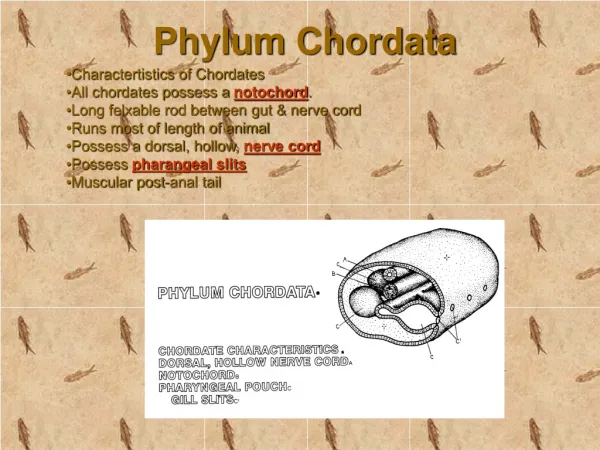
Slide 2 What is Neocallimastigomycota? Anaerobic fungi belong to the phylumNeocallimastigomycota, the earliest diverging lineage unequivocally assigned to kingdom Fungi and are closely related to the chytrids (phylum Chytridiomycota). While they share key morphological features with their chytrid relatives, they possess a distinctive anaerobic physiology and flagellar apparatus. Furthermore, genetic analyses have consistently shown that the Neocallimastigo-mycotaform a distinct, well-supported clade basal to the chytrids.
Slide 3 This Anaerobic fungi were originally placed within phylum Chytridio-mycota, within Order Neocallimastigalesbut later raised to phylum level, a decision upheld by later phylogenetic reconstructions. It encompasses only one family. The phylum member living as symbionts that found in the digestive tracts of larger herbivores.The fungi in Neocallimastigomycota were first recognised as fungi by Orpin in 1975, based on motile cells present in the rumen of sheep. Their zoospores had been observed much earlier but were believed to be flagellate protists, but Orpin demonstrated that they possessed a chitin cell wall.
Slide 3 Since their discovery they have been isolated from the digestive tracts of over 50 herbivores, including ruminant and non-ruminant (hindgut-fermenting) mammals and herbivorous reptiles. The phylum Neocallimastigomycota currently comprises six genera, each distinguishable by morphological features: thallus morphology (rhizoidal vs. bulbous) and zoospore flagellation (monoflagellate vs. polyflagellate).
Slide 3 Table 1 Morphological classification of the currently recognised anaerobic fungal genera
Fig. 1 Phase contrast (a–d) and bisbenzimide stained fluorescence (e and f) microscopy images of various anaerobic ... FEMS Microbiology Ecology, Volume 90, Issue 1, October 2014, Pages 1–17, https://doi.org/10.1111/1574-6941.12383 The content of this slide may be subject to copyright: please see the slide notes for details.
Slide 3 • Fig. 1 Phase contrast (a–d) and bisbenzimide stained fluorescence (e and f) microscopy images of various anaerobic fungal morphological features (scale bar, 50 μm): (a) free polyflagellated zoospores in a mixed culture; (b) germination and rhizoidal development of a monocentric, filamentous Piromyces sp.; (c) Polycentric sporangia of an Anaeromyces sp.; (d) bulbous rhizoidal system of Caecomyces sp.; (e) nuclear migration to rhizoids (white arrow) in a polycentric isolate; (f) nucleated rhizoids of Orpinomycesjoyonii. • Unless provided in the caption above, the following copyright applies to the content of this slide: © 2014 Federation of European Microbiological Societies. Published by John Wiley & Sons Ltd
Fig. 2 Profile Neighbour Joining tree of NeocallimastigomycotaITS1 sequences based on 575 unique ITS1 sequences ... FEMS Microbiology Ecology, Volume 90, Issue 1, October 2014, Pages 1–17, https://doi.org/10.1111/1574-6941.12383 The content of this slide may be subject to copyright: please see the slide notes for details.
Slide 3 • Fig. 2 Profile Neighbour Joining tree of NeocallimastigomycotaITS1 sequences based on 575 unique ITS1 sequences across the Neocallimsatigomycota. The predicted secondary structure of the ITS1 region was modelled to generate an improved alignment of sequences. In addition to the six named genera, it is apparent that ten or more clades, which at present are unnamed, also exist. This figure has been reprinted with permission from Koetschan et al. (2014).
Slide 3 Life cycle Anaerobic fungi reproduce through the asexual production of flagellated zoospores from sporangia, with no sexual reproductive life stage identified to date. Zoospores may be posteriorly monoflagellate or polyflagellate, with the subsequently developed thallus being either monocentric (single reproductive body i.e. one sporangium from single zoospore) or polycentric (many sporangia from a single zoospore). In the rumen, zoospores are released from anaerobic fungal sporangia in response to ingestion of food, with the timing of peak zoospore density being reached within 30–60 min. Ruminal zoospore differentiation and subsequent maturation are thought to occur through the induction of sporangia by haem and other related porphyrins (Fig.), which are released from ingested plant material.
Slide 3 Life cycle Fig. _. Summary of the anaerobic fungal life cycle. The stages in the life cycle where ‘resistant’ structures (that have been reported to date) may be formed are also indicated (*).
Slide 3 Reproduction and growth These fungi reproduce in the rumen of ruminants through the formation of zoospores which are released from sporangia. These zoospores bear a kinetosome but lack the nonflagellated centriole known in most chytrids, and have been known to utilize horizontal gene transfer in their development of xylanase (from bacteria) and other glucanases. The nuclear envelopes of their cells are notable for remaining intact throughout mitosis. Sexual reproduction has not been observed in anaerobic fungi. However, they are known to be able to survive for many months in aerobic environments, a factor which is important in they colonisation of new hosts. In Anaeromyces, the presence of putative resting spores has been observed but the way in which these are formed and germinate remains unknown.
Slide 3 Metabolism & Polysaccharide-degrading activity Metabolism Neocallimastigomycotalack mitochondria but instead contain hydrogenosomes in which the oxidation of NADH to NAD+, leading to formation of H2. Polysaccharide-degrading activity Neocallimastigomycota play an essential role in fibre-digestion in their host species. They are present in large numbers in the digestive tracts of animals which are fed on high fibrediets. The polysaccharide degrading enzymes produced by anaerobic fungi can hydrolyse the most recalcitrant plant polymers and can degrade unlignified plant cell walls entirely. The polysaccharide degrading enzymes are organised into a multiprotein complex, similar to the bacterial cellulosome.
Slide 11 THANK YOU