Corynebacterium & Listeria
290 likes | 565 Vues
Corynebacterium & Listeria. Dr. Zaheer Ahmed Chaudhary Associate Professor Microbiology Department of Pathology. Corynebacterium. Morphology. Club shaped Gram positive rods L-V formation (Chinese letters) Beaded appearance containing highly polymerised Polyphoshate

Corynebacterium & Listeria
E N D
Presentation Transcript
Corynebacterium & Listeria Dr. Zaheer Ahmed Chaudhary Associate Professor Microbiology Department of Pathology
Morphology • Club shaped Gram positive rods • L-V formation (Chinese letters) • Beaded appearance containing highly polymerisedPolyphoshate • Granules stained metachromatically • Aerobic, facultative anaerobe. • Non sporing, non capsulated.
Types of Bacteria • Corynebacterium Diphtheriae • Corynebacterium Ulcerans • Corynebacterium Haemolyticum • Corynebacterium Ovis • Corynebacterium Pyogenes • Corynebacterium Xerosis • Corynebacterium Hofmani • JK Group
Mode of Transmission • Humans are natural host of C. diphtheriae (toxigenic and non toxigenic) • Resides in the upper respiratory track and skin. • Transmitted by air-born droplets • Poor skin hygiene and indigent persons are the victims.
Normal Habitat • Soil, plants and animals • In humans, commensal dipheroids form part of the skin flora, upper respiratory tract, urinary tract and conjunctiva.
Bio Types of C.diphtheriae • Based on the severity of infections, there are 3 bio types • Gravis • Mitis • Intermedius
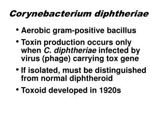
Pathogenesis • Powerful exotoxin is produced. • Organism get attached to the throat epithelium. • Diphtheria toxin inhibits protein synthesis by ADP ribosylation of elongation factor 2. • Temperate bacteriophage • The toxin causes local inflammation in the throat with fibrinous exudate that forms tough, adherent grey pseudomembrane.
Clinical Features • Thick, grey-white adherent pseudomembrane over tonsils and throat. • Fever, sore throat, cervical lymphoadenopathy.
Complications • Extension of pseudomembrane into Larynx and trachea causing air-way obstruction and death. • Myocarditis with arrhythmias and circulatory collapse. • Nerve weakness and paralysis of cranial nerves. • Paralysis of muscles of soft pallet and pharynx leading to regurgitation of fluids through nose. • Peripheral neuritis affecting the muscles of extremities.
Lab Diagnosis • Purely clinical diagnosis • Isolation of the organism • Toxin production demonstration • Schick’s Test
Culture • Does not grow on ordinary media • Special media • Lofflers medium • Tellurite medium • Typically grey-black color of Tellurium in the colony is diagnostic. • Gram stain shows tapperedpleomorphic rods • Methylene blue stained shows metachromatic granules.
Treatment • Anti-toxin – neutralizes the unbound toxin in the blood. • Hypersensitivity test • Penicillin G, erythromycin
Vaccination • Diphtheria toxoid – 3 doses (2,4,6 years) • Booster dose after every 10 years
Characteristics • Small gram positive rods arranged in v and L arrangement. • Motile with tumbling movements. • Growth on blood agar plate produces narrow zone of B-hemolysis. • Growth at low temperature e.g. cold storage and refrigerator. • Causes meningitis and sepsis in new born, pregnant women and immunocompromised adults. • Occasionally it causes outbreaks of gastroenteritis.
Pathogenesis • Can produce diseases in • Fetus • Pregnant women and immunocompromised adults. • Organism has world wide distribution and is present in animals, soil and plants. • Human get infected by ingestion of unpasteurized milk, uncooked meat and raw vegetables.
Contacts with pets and their feces can also be an important source. • After ingestion the bacteria reach the colon and later colonize the female genital tract. Fetus can get infected after rupture of membranes while passing through the birth canal. • Invasion of the body is mediated by internalin protein (produced by listeria) and E. cadherin on the surface of human cells.
The organism produces listeriolysin on entering the cell which allows it to escape from phagosome and escape its destruction. • Listeria is intracellular bacteria and cell mediated immunity plays major role is host defense • Suppression of cell mediated immunity predispose to listera infections.
Clinical findings • Infection during pregnancy can result in abortion, pre mature delivery or sepsis • New born infected can have acute meningitis after 1 – 4 weeks • Infected mother can be asymptomatic or have flue like symptoms. Immunocompromised adults can have sepsis or meningitis, • Gastroenteritis can present as watery diarrhea, fever, headache, myalgia and little vomiting.
Outbreaks can occur by eating contaminated dairy products, uncooked meat, ready to eat food like Cole-slaw etc.
Lab diagnosis • Gram stain • Culture • Motility • Sugar fermentation test
Treatment • Septran • Ampicillin and gentamycin
Prevention • Limiting the exposure of pregnant women and immunocompromised patients to potential dangerous sources like farmhouses, unpasteurized milk products and raw vegetables.