Download
1 / 32
330 likes | 371 Vues
Explore the intricate development of arteries and veins in fetal circulation, from vitelline veins to cardinal veins and more. Understand the role of different veins and the unique pathways they follow during embryonic growth.

E N D
Development of the arteries and veinsFetal circulation Mark Kozsurek, M.D., Ph.D. mark@kozsurek.hu http://gut.bmj.com/content/57/7/F1.medium.gif ED I., 20/02/2018
1 2 3
sinus venosus atria ventricles anterior aspect !!!
anterior cardinal vein common cardinal vein cut edge ot the sinu-atrial junction sinus venosus umbilical vein vitelline vein posterior cardinal vein
anastomosis between the right and left anterior cardinal veins anterior cardinal vein sinus venosus posterior cardinal vein
anastomosis between the right and left anterior cardinal veins anterior cardinal vein sinus venosus posterior cardinal vein anastomosis between the right and left posterior cardinal veins
right and left brachiocephalic veins superior vena cava oblique vein of the left atrium coronary sinus sinus venosus posteriorcardinal vein right and left common iliac veins
DUODENUM right and left brachiocephalic veins superior vena cava oblique vein of the left atrium coronary sinus sinus venosus posteriorcardinal vein right and left vitelline veins from the yolk sac right and left common iliac veins
right and left brachiocephalic veins superior vena cava oblique vein of the left atrium coronary sinus sinus venosus posteriorcardinal vein right and left vitelline veins from the yolk sac DUODENUM right and left common iliac veins
right and left brachiocephalic veins superior vena cava oblique vein of the left atrium coronary sinus sinus venosus right vitelline vein posteriorcardinal vein DUODENUM right and left common iliac veins
right and left brachiocephalic veins superior vena cava oblique vein of the left atrium coronary sinus sinus venosus right vitelline vein posteriorcardinal vein DUODENUM right and left common iliac veins
right and left brachiocephalic veins superior vena cava oblique vein of the left atrium coronary sinus sinus venosus right vitelline vein posteriorcardinal vein left umbilical vein placenta umbilical veins right and left common iliac veins
supracardinal vein subcardinal vein kidney right and left brachiocephalic veins superior vena cava oblique vein of the left atrium coronary sinus sinus venosus placenta right and left common iliac veins
liver right and left brachiocephalic veins superior vena cava oblique vein of the left atrium coronary sinus sinus venosus supracardinal vein subcardinal vein placenta kidney right and left common iliac veins
ductus venosus liver right and left brachiocephalic veins superior vena cava oblique vein of the left atrium coronary sinus sinus venosus supracardinal vein subcardinal vein placenta kidney right and left common iliac veins
ductus venosus liver right and left brachiocephalic veins superior vena cava oblique vein of the left atrium coronary sinus sinus venosus supracardinal vein subcardinal vein placenta kidney right and left common iliac veins
hemiazygos vein accessory hemiazygos vein azygos vein portal vein splenic vein superior mesenteric vein gonadal veins sinus venosus liver placenta
In the early stage of the embryonic development a pair of vitelline veins carry blood from the yolk sac, but later only the right one persists. • With the appearance and progressive development of the placenta the role of the vitelline veins is replaced by a pair of umbilical veins, but only the left one remains between the placenta and the primordial heart. • The main venous system draining the head and the caudal part of the embryo is represented by the anterior and posterior cardinal veins, respectively. • Supracardinal veins appear on the lateral side of the developing kidney. The supracardinal veins are connected with a horizontal anastomosis, resulting in a H-shaped structure.
On the medial sides of the primordial kidneys a new pair of veins develop: the subcardinal veins, which are also interconnected with a horizontal anastomosis. • The liver developing in the septum transversum devides the remaining right vitelline vein in three segments: prehepatic, intrahepatic and posthepatic parts can be distinguished. • Due to the growing liver the left umbilical vein is interrupted and looses its contact with the heart. The larger proportion of the blood arriving from the placenta enters the inferior vena cava on the surface of the liver via the ductus venosus, while the rest enters the hepatic sinuses.
On the left side the cardinal veins degenerate. The only notable persisting vessel is the coronary sinus. • The right anterior cardinal vein develops into the right brachiocephalic vein and the superior vena cava, while the anastomosis between the two anterior cardinal veins remains as the left brachiocephalic vein. • Supra- and subcardinal veins with the prehepatic segment of the right vitelline vein and the interconnecting anastomoses play an important role in formation of the inferior vena cava. • Prehepatic segment of the right vitelline vein develops into the portal vein. • The major part of the supracardinal veins persists as the vena azygos/hemiazygos system. • Subcardinal veins are incorporated into the inferior vena cava or develop into the renal and gonadial veins.
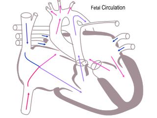
Fetal circulationPostnatal adaptation of the circulatory system
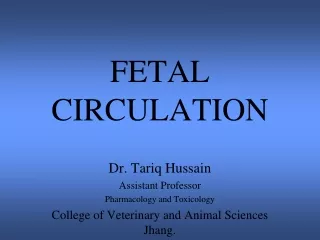
Arterious blood is carried into the fetus by the umbilical vein. A smaller proportion of this blood passes through the liver and is collected by the IVC. The majority of the oxygeneted blood bypasses the liver through the ductus venosus and drains directly into the IVC. The Eustachian valve directs the blood toward the foramen ovale and the left atrium. This blood is then pumped into the aorta and supplies the tissues of the fetus. Finally returnes to the placenta via the two umbilical erteries arising from the internal iliac arteries. • Venous blood derived from the head and neck regions as well as from the upper limb is collected by the SVC. This venous blood gets into the right atrium then into the right ventricle. As the lungs are collapsed the vascular resistance is very high: the blood may not flow toward the pulmonary arteries. This is why the ductus arteriosus is essential: it drains the venous blood of the pulmonary trunk into the aorta.Briefly: during the fetal period both of the ventricles eject blood into the aorta!!!
After birth no more arterious blood arrives through the umbilical vein. The lungs are inflated, the vascular resistance suddenly drops. There is no further reason for the blood ejected by the right ventricle to join the aorta. As more blood reaches the lungs, more returns through the pulmonary veins into the left atrium. The pressure increases here and the septum primum is pushed against the septum secundum: the foramen ovale closes, later the two septa completly fuse. • Due to the increased oxygen levels smooth muscles of the ductus arteriosus contract and obliterate the lumen. (Otherwise the higher pressure in the aorta would result in a reversed flow through the ductus venosus.) • Ductus venosus is slowly occupied by proliferating connective tissue and remains observable on the visceral surface of the liver as the venous ligament.