Gallbladder Carcinoma
Gallbladder Carcinoma. SONO 1218 March, 2008. Gallbladder Carcinoma. Although uncommon, carcinoma of the gallbladder is the most common primary hepatobiliary carcinoma. When the diagnosis is made incidentally at the time of cholecystectomy, surgical resection can be curative.

Gallbladder Carcinoma
E N D
Presentation Transcript
Gallbladder Carcinoma SONO 1218 March, 2008
Gallbladder Carcinoma • Although uncommon, carcinoma of the gallbladder is the most common primary hepatobiliary carcinoma. • When the diagnosis is made incidentally at the time of cholecystectomy, surgical resection can be curative. • More commonly, the tumor is unresectable and rarely diagnosed preoperatively despite patients' symptoms.
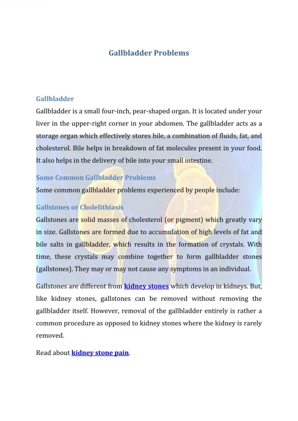
Gallbladder Carcinoma • How It SpreadsGall bladder cancer tends to spread to nearby organs and tissues. (i.e. liver or small intestine). It is also spread through the lymphatic system. • What Causes ItNo one factor has been clearly shown to cause gallbladder cancer. • It is more often seen in older patients with long-standing cholecystolithiasis. • Common Signs and SymptomsThere are no firm clinical signs or symptoms characteristic of gallbladder cancer. • Jaundice (the skin turning yellow), bloating, abdominal pain, weight loss, decreasing appetite, fever, nausea or an enlarging abdominal mass can all be attributable to gallbladder cancer. Even if these symptoms are found gall bladder cancer would still not be immediately suspected because it is so uncommon.
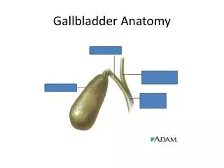
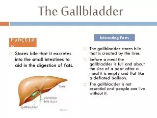
Ultrasound and Gallbladder Carcinoma • Ultrasound studies have demonstrated the gall bladder wall to consist of two or more (usually three) layers. • Gall bladder tumors are usually confined to the mucosa or muscle layer, or have only invaded the shallow subserosa.
Our Patient • An apparently healthy 53-year-old woman with recurrent epigastric pain • She was found to have a 2.5cm stone in the gallbladder • Additionally, there was a hypoechoic mass in the fundal region that measured 3.3 X 2.9cm
Gallbladder Carcinoma • This image shows the gallstone • It appears to be isolated in this view
This image still shows the stone but the shadowing posterior to the the fundal area clearly indicates an area of concern
Gallbladder Carcinoma • The hypoechoic mass measured 3.3 X 2.9cm
Gallbladder Carcinoma The image shows the thickened wall of the gallbladder
Gallbladder Carcinoma Because of the lack of blood flow to the mass the diagnosis was not made via Ultrasound but was still pending hystology results
CREDITS • http://www.cancersupportivecare.com/gallbladder.html • http://www.ultrasound-images.com/gall-bladder.htm