LIVER& GALLBLADDER
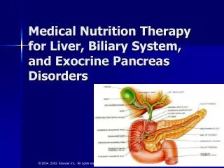
LIVER& GALLBLADDER. Dr Iram Tassaduq. Major salivary glands Gallbladder Pancreas Liver. LIVER. LIVER. Largest mass of glandular tissue Largest internal organ 1500 g in weight. ANATOMY OF LIVER. STRUCTURAL ORGANIZATION. Parenchyma Connective tissue stroma

LIVER& GALLBLADDER
E N D
Presentation Transcript
LIVER&GALLBLADDER Dr Iram Tassaduq
Major salivary glands • Gallbladder • Pancreas • Liver
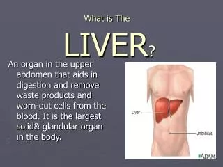
LIVER • Largest mass of glandular tissue • Largest internal organ • 1500 g in weight
STRUCTURAL ORGANIZATION Parenchyma Connective tissue stroma Sinusoidal capillaries (sinusoids) Perisinusoidal space (space of Disse)
CAPSULE • The liver is enclosed in a capsule of fibromuscular connective tissue known as Gilson's capsule. • Thin capsule
LIVER LOBULES • Classic Lobule • Portal Lobule • Liver acinus
PORTAL TRIAD • Hepatic artery • Portal vein • Bile duct
Portal lobule • Emphasizes exocrine function • Includes those portions of three classical lobules that secrete bile that drains unto bile duct
FORMATION OF LYMPH IN THE LIVER • Due to the large pores or fenestrations in sinusoidal endothelial cells, fluid and proteins in blood flow freely into the space between the endothelium and hepatocytes (the "space of Disse"), forming lymph. Lymph flows through the space of Disse to collect in small lymphatic capillaries associated with portal triads.
SPECIAL FEATURES OF MUCOSA • Microvilli • Junctional complexes • Concentration of mitochondria • Lateral plications