Download
1 / 81
1.03k likes | 2.73k Vues
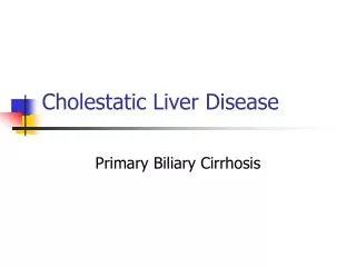
Cholestatic Liver Disease - PBC and PSC. Cholestasis: Definition. Cholestasis implies that the liver is not producing or secreting bile normally Biochemically: Elevated alkaline phosphatase, conjugated bilirubin or both Signs/symptoms: Pruritus Jaundice Histologically Bile lakes

E N D
Cholestasis: Definition • Cholestasis implies that the liver is not producing or secreting bile normally • Biochemically: • Elevated alkaline phosphatase, conjugated bilirubin or both • Signs/symptoms: • Pruritus • Jaundice • Histologically • Bile lakes • Cholate stasis • Ductular proliferation
Cholestasis: Definition • Cholestasis can be divided into 2 basic categories: • Extrahepatic Obstructive Jaundice • Intrahepatic Cholestasis
Extrahepatic Obstructive Cholestasis • Extrahepatic obstruction of the bile ducts results in upstream ductal dilation • Seen on imaging • The primary considerations are: • pancreatic cancer • cholangiocarcinoma • choledocholithiasis • benign papillary stenosis
Intrahepatic Cholestasis • Direct effect on cannalicular membrane of the hepatocyte by: • Toxins, metabolites • Drugs, alcohol • Interleukins, tumor necrosis factor • ALP (a hepatocellular enzyme) translocates to the basolateral membrane and is lost to serum
Intrahepatic Cholestasis: Etiologies • Drugs • OCPs, anabolic steroids, NSAIDs, antibiotics (e-mycin), antipsychotics • Infiltrative disorders • Lymphoma, amyloidosis, tuberculosis, sarcoidosis, metastases • TPN • Sepsis • Pregnancy • Autoimmune (PBC/PSC, AIH overlap syndrome) • Inherited (benign recurrent intrahepatic cholestasis) • Post-operative • Paraneoplastic (Stauffer’s syndrome)
Alkaline Phosphatase • ALP is a protein located on the cannalicular membrane of the hepatocyte – NOT the bile duct cell • Widespread tissue distribution: • Liver • Bone • Intestine • Kidney • placenta
Alkaline Phosphatase • In biliary obstruction, increased synthesis and release of the enzyme into the serum rather than impaired biliary secretion accounts for the elevation of ALP • In cholestasis, bile acids accumulate in the hepatocytes and solubilize the plasma membrane resulting in release of ALP into serum • The level not a reliable indicator of the severity of underlying liver disease
Alkaline Phosphatase:Confirming Liver Source • Gamma-glutamyl transferase (GGT) • Can be isolated from hepatocytes and various extrahepatic tissues but NOT bone • 5’ Nucleotidase • Elevated 5NT is rarely associated with associated with an extrahepatic source • Fractionate the alkaline phosphatase • Any elevation in a transaminase is very specific for liver origin of ALP
Primary Biliary Cirrhosis:Overview • Definition • Epidemiology • Pathogenesis • Natural history • Clinical features • Diagnosis • Pathology • Management • Complications • Transplantation
Primary Biliary Cirrhosis:Definition • Chronic cholestatic liver disease • Hallmark serologic signature – the antimitochondrial antibody (AMA) • Chronic, non-suppurative destructive cholangitis on histology
Primary Biliary Cirrhosis:Epidemiology • Worldwide • Predominantly women (9:1) • Diagnosed typically between ages 30-60 • In the United States: • Age adjusted incidence rate is 27/million person-years • Age adjusted prevalence is 402/million
Primary Biliary Cirrhosis:Pathogenesis • Cause is unknown • Considered an autoimmune disorder • Intense humoral and cellular response to an intracytoplasmic antigen • Presence of highly specific autoantibodies • Involvement of T lymphocytes in the destruction of bile ducts • Often coexists with other autoimmune disorders
Proposed Mechanisms for the development of PBC • Molecular mimicry (Microorganism infection) • Xenobiotics • Genetic
Primary Biliary Cirrhosis:Natural History Springer et al. Am J Gastro 1999
Primary Biliary Cirrhosis:Natural History Springer et al. Am J Gastro 1999
Primary Biliary Cirrhosis:Natural History • In general, slowly progressive disorder • Consists of two phases: • Asymptomatic: • Lasting mean of 17 years • No liver related mortality during this time • Symptomatic: • Characterized by the onset of symptoms • Average time from symptom onset to death is 7 years
Primary Biliary Cirrhosis:Natural History Predicting Survival – The Mayo Risk Score • In general, if bilirubin is > 10 there is a 50% survival at 2 years
Primary Biliary Cirrhosis:Clinical Features at Presentation • Asymptomatic 40-60% • Fatigue 21-85% • Pruritus 19-55% • Sicca symptoms up to 70% • Hepatomegaly 25% • Splenomegaly 15% • Jaundice uncommon • Xanthelasma uncommon
Primary Biliary Cirrhosis:Other Associated Conditions • Metabolic bone disease • Fat soluble vitamin deficiencies • Hypercholesterolemia • Autoimmune thyroiditis • CREST/Raynaud’s • Renal tubular acidosis • Celiac sprue
Primary Biliary Cirrhosis:Diagnosis • Elevated alkaline phosphatase • Typically 3-4 X ULN • ALT/AST usually < 200 • Unremarkable biliary imaging • Elevated immunoglobulins • Typically IgM • Characteristic autoantibody (AMA) • Characteristic histology
Primary Biliary Cirrhosis:Diagnosis Autoantibodies: • Antimitochondrial Antibodies (AMA) • Present in 95% of patient with PBC • Sensitivity and specificity are > 95% • Usually present in high titer • Antinuclear/Smooth Muscle Antibodies (ANA/ASMA) • Not specific • Seen in 1/3 to ½ of individuals with PBC
Primary Biliary Cirrhosis:Liver Biopsy Staging: Ludwig’s or Scheuer’s Classification Stage 1 portal inflammation/florid duct lesion Stage 2 periportal inflammaton (interface hepatitis), ductular proliferation Stage 3 bridging fibrosis, ductopenia Stage 4 cirrhosis
Histological Staging – PBC Primary Biliary Cirrhosis – Histological Features Stage 1 Stage 2 Stage 3 Stage 4
Histological Staging – PBC – Stage 1 Primary Biliary Cirrhosis Stage 1: Florid Duct Lesion Granuloma Portal hepatitis Scheuer P, Proc R Soc Med 1967; 60:1257 Ludwig, et al., Virch Arch Pathol Anat 1978; 379:103
Histological Staging – PBC – Stage 2 Primary Biliary Cirrhosis Stage 2: Ductular Reaction Periportal Hepatitis Scheuer P, Proc R Soc Med 1967; 60:1257 Ludwig, et al., Virch Arch Pathol Anat 1978; 379:103
Histological Staging – PBC – Stage 3 Primary Biliary Cirrhosis Stage 3: Scarring Bridging Necrosis/ Septal Fibrosis Scheuer P, Proc R Soc Med 1967; 60:1257 Ludwig, et al., Virch Arch Pathol Anat 1978; 379:103
Histological Staging – PBC – Stage 4 Primary Biliary Cirrhosis Stage 4: Cirrhosis Cirrhosis Scheuer P, Proc R Soc Med 1967; 60:1257 Ludwig, et al., Virch Arch Pathol Anat 1978; 379:103
Primary Biliary Cirrhosis:Treatment • Corticosteroids • Colchicine • Azathioprine • Cyclosporine • Methotrexate
Primary Biliary Cirrhosis:Treatment • Corticosteroids • Colchicine • Azathioprine • Cyclosporine • Methotrexate
Primary Biliary Cirrhosis:Treatment • UDCA in a dose of 13-15 mg/kg/day is the only FDA approved therapy for PBC • Dose is important • Number of studies have demonstrated a clear benefit • Used in all stages of PBC
Primary Biliary Cirrhosis:Actions of UDCA • Protects against cytotoxic effects of di-hydroxy bile acids • Modulates expression of HLA • Stabilizes bile canalicular membrane • Choleretic effect • Decreased apoptosis • Decreased cytokine production
Nonresponder Responder Pares et al. Gastro 2006
Primary Biliary Cirrhosis:Management • Liver chemistries every 3-6 months • Thyroid study (TSH) annually • Bone mineral density scan (DEXA) every 2-4 years • Fat soluble vitamin levels if bilirubin > 2 • Upper endoscopy if cirrhotic or Mayo risk score > 4.1 • Ultrasound +/- AFP every 6-12 months if cirrhotic
Primary Biliary Cirrhosis:Management Fatigue: • Modafanil 100-200mg/d Pruritus: • Cholestyramine up to 4g qid • Rifampicin 150mg/d • Naltrexone 12.5 mg/d (up to 50mg/d) Sicca: • Artificial tears, saliva substitutes • Good dental hygiene • Pilocarpine or cevimeline Osteopenia/Osteoporosis: • 1000-1500mg/d calcium + 1000 IU Vit D • Bisphosphanates Hyperlipidemia: • No treatment, no increased risk of cardiovascular disease
Primary Biliary Cirrhosis:Transplantation • PBC was the leading indication for OLT in the U.S. in the mid 1980s • Despite increased numbers of transplants, patients with PBC requiring OLT have decreased by 20% • Outcome of OLT in PBC is more favorable than almost all other liver diseases • Once listed, priority is based on MELD