Cell Communication and Signaling Mechanisms in Yeast and Animals
Explore the intricate pathways of cellular communication and signaling in yeast and animals, including mechanisms such as G-protein-linked receptors and phosphorylation cascades. Understand the role of second messengers like cAMP and the specificity of cell signaling processes.

Cell Communication and Signaling Mechanisms in Yeast and Animals
E N D
Presentation Transcript
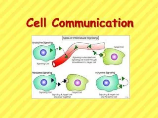
Figure 11.3 Local and long-distance cell communication in animals
Figure 11.8 The structure and function of a tyrosine-kinase receptor
Figure 11.10 Steroid hormone interacting with an intracellular receptor
Figure 11.14 The maintenance of calcium ion concentrations in an animal cell
Figure 11.15 Calcium and inositol triphosphate in signaling pathways (Layer 3)
Figure 11.16 Cytoplasmic response to a signal: the stimulation of glycogen breakdown by epinephrine
Figure 11.17 Nuclear response to a signal: the activation of a specific gene by a growth factor
Figure 12.3 Chromosome duplication and distribution during mitosis
Figure 12.5 The stages of mitotic cell division in an animal cell: G2 phase; prophase; prometaphase
Figure 12.5 The stages of mitotic cell division in an animal cell: metaphase; anaphase; telophase and cytokinesis.
Figure 12.7 Testing a hypothesis for chromosome migration during anaphase
Figure 12.10 Bacterial cell division (binary fission) (Layer 3)
Figure 12.17 The growth and metastasis of a malignant breast tumor
Figure 12-17x2 Mammogram: normal (left) and cancerous (right)
Figure 13.x2 Human female chromosomes shown by bright field G-banding
Figure 13.x3 Human female karyotype shown by bright field G-banding of chromosomes