Download
1 / 25
250 likes | 278 Vues
This case study details a unique dentigerous cyst in a 5-year-old Paint gelding, including symptoms, differential diagnoses, diagnostic procedures, and treatment plan, shedding light on this uncommon equine condition.

E N D
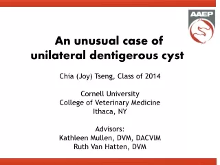
An unusual case of unilateral dentigerous cyst Chia (Joy) Tseng, Class of 2014 Cornell UniversityCollege of Veterinary MedicineIthaca, NY Advisors: Kathleen Mullen, DVM, DACVIM Ruth Van Hatten, DVM
SIGNALMENT • 5-year-old Paint gelding • Purchased 1.5 years ago from Iowa • No prior medical history except for hard swellingat base of right ear of unknown duration • Intended use: show horse • Sire: HYPP double negative
HISTORY Mar 2014 • R facial nerve paralysis • Vestibular episode post-sedation • Skull radiograph: R dentigerous cyst Jan 2014 • Excessive head shaking • Swelling at base of right ear • FNA of swelling: • Cartilaginous material • No infection • Phenylbutazone • Flunixin meglumine • No improvement 2 weeks later • Acute lethargy • Acute uncoordination • Right droopy lip • Oral exam: normal • Neuro exam: normal • Corneal ulcer OD • Trimethoprim/sulfadiazine x 14 days • Minimal improvement
INITIAL EXAM- CORNELL • Bright, alert, responsive, normal mentation • T 101F HR 48/min RR 24/min • No murmur, arrythmias, or adventitious lung sounds • Mucous membrane pink and moist, CRT <2s • Centroventral corneal opacity OD • Fundic exam normal OU
INITIAL EXAM Cont’d • Good anal and tail tone • Erect and fixed R ear • 5cm x 5cm firm swelling at base of R pinna • All other body systemshad no abnormality
PROBLEM LIST • R facial nerve paralysis CN VII • Scar from presumed secondary exposure keratitis OD • Vestibular signs CN VIII • Erect and fixed R pinna • Presumptive R dentigerous cyst
DDx • Firm swelling at base of R pinna: • Dentigerous cyst • Neoplasia (Osteoma, Ameloblastoma) • Calcified granuloma • Abscess • Hematoma • DDx for acute CNs VII and VIII signs: • Peripheral diseases • Temporohyoid osteoarthropathy • Otitis interna/media • Rarely guttural pouch inflammation/infection • Central vestibular disease ruled out • No mentation change • No hypermetric gait abnormality • No vertical nystagmus • No paradoxical vestibular signs
DIAGNOSTIC PLAN • Head radiographs- performed by rDVM • Endoscopic exam of guttural pouches • Computed tomography (CT) of head • General anesthesia required • Surgical planning
HEAD RADIOGRAPH Rostral Rostral RLAT RLAT • Mineral opacity at base of the pinna dorsal to the guttural pouch • No fluid line in guttural pouch
ENDOSCOPY • Right guttural pouch: • Mass* protruding into dorsolateral compartment • Concretion-like material in dorsomedial compartment* • Diffusely inflamed mucosa • Mobile hyoid apparatus when basihyoid was palpated • Movement of mass when right ear manipulated • Left guttural pouch: normal * stylohyoid R-GP: dorsolateral * stylohyoid R-GP: dorsomedial
HEAD CT • An amorphous, mineralized mass resembling multiple teeth at the R temporal bone • Protruding into dorsal aspect of R guttural pouch • Occluding R external ear canal • R otitis externa, media, interna • Normal R temporohyoid joint • No contrast communication between mass and concretion R Transverse: level of tympanic bulla R Transverse: level of petrous temporal bone
DDx REVISITED • Firm swelling at base of R pinna: • Dentigerous cyst presumptive • Neoplasia (Osteoma, Ameloblastoma) • Calcified granuloma • Abscess • Hematoma • DDx for acute peripheral CNs VII and VIII signs: • Temporohyoidosteoarthropathy • Otitis media/interna • Rarely guttural pouch inflammation/infection • Right GP mass • Dentigerous cyst • Hematoma • Neoplasia • Right GP concretion (Bacterial culture submitted) • Abscess • Fungal plaque • Neoplasia
DIAGNOSIS • Right dentigerous cysts • Secondary otitis externa, media and interna • Chronic lack of drainage through auditory tube due to dentigerous cyst blockage • Secondary Streptococcus equi ssp. zooepidemicus infection
CULTURE & SENSITIVITY • Streptococcus equi ssp. zooepidemicus:sample taken sterilely from lateral compartment in right guttural pouch Highlighted: oral formulation available
DEVELOPMENT OF THE FACE Branchial arches • By 4 weeks, pharyngeal pouches develop ventral to the pharynx • Neural crest cells segregate into a series of branchial arches • Mesenchymal cells within the 1st branchial arch grow in 2 directions to form the maxillary and mandibular processes • Odontogenic cells migrate into the 1st branchial arch to form teeth within the jaws Frontonasalprominence Lateral view Maxillary process Mandibular process 2nd branchial arch Ventral view Approx. 25-day old embryo diagram Modified from The Embryology of Domestic Animals 1985
DENTIGEROUS CYST • Etiology: • Failure of odontogenic cells to migrate fixed in improper location • 1st branchial arch fails to close fistulous tract • Abnormal tooth with cystic lining displaced towards the temporal region • Reported locations: • Temporal region- most common (Easley et al. 2010) • Cyst adhered to calvarium (Easley et al. 2010) • Adjacent to cerebellum (Hunt et al. 1991) • Ventral nasal meatus (de Mira et al. 2007) • Ventral conchal sinus (McClure et al. 1993)
DENTIGEROUS CYST • Typical clinical manifestation: • 3-5 years old • No breed or sex predilection • Firm, non-painful swelling at base of ear with a fistulous draining tract • Owners typically notice matted hair around draining tract • +/- Secondary bacterial infection • Unilateral > bilateral Photo from a different casecredit: Dr. Ryland Edwards III
DENTIGEROUS CYST • Diagnosis: • Head radiograph • May be hard to interpret but often diagnostic • CT • Higher sensitivity • Important for surgical planning • Treatment: • Surgical excision under general anesthesia usually for cosmetic reason • Lateral approach- complete excision of entire cyst with epithelial lining Photo from a different casecredit: Dr. Ryland Edwards III
OUR UNIQUE CASE • No external draining tract • Acute onset facial nerve paralysis & peripheral vestibular signs • Dentigerous cyst protrudes into roof of guttural pouch • Secondary bacterial otitis externa, media, interna • Internal abscessation exiting into the guttural pouch
OUR TREATMENT OPTIONS Surgical removal of dentigerous cyst VS. Medical management of otitis first * Resolution of vestibular signs is UNKNOWN with either option *
DECISION MAKING • Medical management of otitis was recommended and elected • Location of the dentigerous cyst requires highly extensive surgery with many potential complications due to location near several major cranial nerves and vessels • High risk of recovery complication in a vestibular patient • Risk of bacterial infection spreading to deeper tissues • Presenting clinical signs are thought to be due to the secondary otitis; therefore, if otitis can be managed medically, a potentially highly invasive and risky surgery can be delayed or avoided • Surgical intervention was recommended IF medical management fails or if otitis and/or vestibular signs recur
OUTCOME • Medical management of otitis with pending surgical option • 30-day course Enrofloxacin (7.5 mg/kg PO q24h) • Chosen for its good tissue penetration among the other available oral antibiotics listed 2-week report • R unilateral nasal drainage of purulent material 1-month recheck • Swelling in dorsolateral compartment of R guttural pouch absent • Mild mobility of the R pinna • Minor R facial nerve paralysis • Vestibular signs resolved • Owners declined surgical intervention at this time
FURTHER READING • de Mira MC, Ragle CA, Gablehouse KB, Tucker RL. Endoscopic removal of a molariform supernumerary intranasal tooth (heterotopic polyodontia) in a horse. JAVMA. 2007; 231(9): 1374-7. • Easley JT, Franklin RP, Adams A. Surgical excision of a dentigerous cyst containing two dental structures. Equine vet. Educ. 2010; 22(6): 275-8. • Hunt RJ, Allen D, Mueller PO. Intracranial trauma associated with extraction of a temporal ear tooth (dentigerous cyst) in a horse. The Cor. Vet. 1991; 81(2): 103-8. • McClure SR, Schumacher J, Morris EL. Dentigerous cyst in the ventral conchal sinus of a horse. Vet. Rad. & Ultras. 1993; 34(5): 334-5. • Smith LCR, Zedler ST, Gestier S, Keane SE, Goodwin W, van Eps AW. Bilateral dentigerous cysts (heterotopic polyodontia) in a yearling Standardbred colt. Equine vet. Educ. 2012; 24(11): 573-8.
ACKNOWLEDGEMENT • Case advisory: • Kathleen Mullen, DVM, DACVIM • Ruth Van Hatten, DVM • Tom Divers, DVM, DACVIM, DACVECC • Pete Scrivani, DVM, DACVR • Norm Ducharme, DVM, MSc, DACVS • Christina Cable, DVM, DACVS • Photography & graphics credits: • Emil Olsen, MRCVS, PhD • Ryland Edwards III, DVM, PhD, DACVS • Noden DM & de Lahunta A. The Embryology of Domestic Animals: Developmental Mechanisms and Malformations. Baltimore: Williams & Wilkins. 1985. pp. 162 & 166. A sincere thank you to all who generously shared their knowledge, photos, and enthusiasm for this interesting case as such manifestation of a dentigerous cyst was not previously reported.