Download
1 / 1
10 likes | 128 Vues
Case of the Day – Thursday MUSCULOSKELETAL. Davis, KW 1 and Smith, SE 2 University of Wisconsin Medical School, Madison, Wisconsin 2. Dept. of Diagnostic Imaging, University of Maryland School of Medicine, Baltimore, MD.

E N D
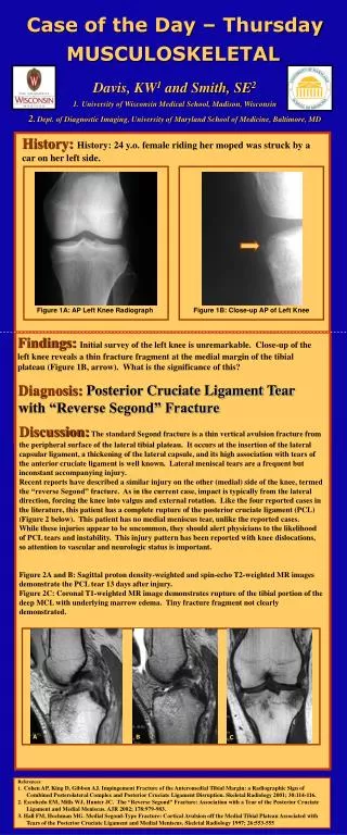
Case of the Day – Thursday MUSCULOSKELETAL • Davis, KW1 and Smith, SE2 • University of Wisconsin Medical School, Madison, Wisconsin • 2. Dept. of Diagnostic Imaging, University of Maryland School of Medicine, Baltimore, MD History:History: 24 y.o. female riding her moped was struck by a car on her left side. Figure 1A: AP Left Knee Radiograph Figure 1B: Close-up AP of Left Knee Findings:Initial survey of the left knee is unremarkable. Close-up of the left knee reveals a thin fracture fragment at the medial margin of the tibial plateau (Figure 1B, arrow). What is the significance of this? Diagnosis: Posterior Cruciate Ligament Tear with “Reverse Segond” Fracture Discussion:The standard Segond fracture is a thin vertical avulsion fracture from the peripheral surface of the lateral tibial plateau. It occurs at the insertion of the lateral capsular ligament, a thickening of the lateral capsule, and its high association with tears of the anterior cruciate ligament is well known. Lateral meniscal tears are a frequent but inconstant accompanying injury. Recent reports have described a similar injury on the other (medial) side of the knee, termed the “reverse Segond” fracture. As in the current case, impact is typically from the lateral direction, forcing the knee into valgus and external rotation. Like the four reported cases in the literature, this patient has a complete rupture of the posterior cruciate ligament (PCL) (Figure 2 below). This patient has no medial meniscus tear, unlike the reported cases. While these injuries appear to be uncommon, they should alert physicians to the likelihood of PCL tears and instability. This injury pattern has been reported with knee dislocations, so attention to vascular and neurologic status is important. Figure 2A and B: Sagittal proton density-weighted and spin-echo T2-weighted MR images demonstrate the PCL tear 13 days after injury. Figure 2C: Coronal T1-weighted MR image demonstrates rupture of the tibial portion of the deep MCL with underlying marrow edema. Tiny fracture fragment not clearly demonstrated. A B C References: 1. Cohen AP, King D, Gibbon AJ. Impingement Fracture of the Anteromedial Tibial Margin: a Radiographic Sign of Combined Posterolateral Complex and Posterior Cruciate Ligament Disruption. Skeletal Radiology 2001; 30:114-116. 2. Escobedo EM, Mills WJ, Hunter JC. The “Reverse Segond” Fracture: Association with a Tear of the Posterior Cruciate Ligament and Medial Meniscus. AJR 2002; 178:979-983. 3. Hall FM, Hochman MG. Medial Segond-Type Fracture: Cortical Avulsion off the Medial Tibial Plateau Associated with Tears of the Posterior Cruciate Ligament and Medial Meniscus. Skeletal Radiology 1997; 26:553-555