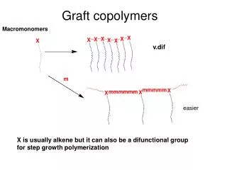
Download
1 / 22
220 likes | 369 Vues
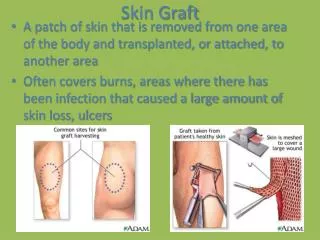
Graft Healing and Return to Play. Joseph F. Scordino September 27, 2007. Case 1.

E N D
Graft Healing and Return to Play Joseph F. Scordino September 27, 2007
Case 1 • The patient was a 35-year-old forward player who sustained an isolated complete tear of the left anterior cruciate ligament (ACL) in the midst of the competitive 2001-2002 season. He was in contention for a position on the Italian World Cup Team that was to be played 135 days after his injury, only if he demonstrated that he could return to play at the highest level before the team was selected. • What graft to use? • What fixation to use? • What is the basic science of graft repair and how does that effect the time to return to sport? • What type of rehab? • How early can he return?
Physiology • Graft serves as scaffold which is rapidly incorporated by the host • Similar to avascular necrosis: cell death to revascularization to cell repopulation to remodeling • Cell Death: first phase in which fibroblasts dye but graft acts as scaffold for new ingrowth. This is an inflammatory stage. • Revascularization: New cells grew into graft. This starts at 20 days and is completed at 6 months. Graft strength in some studies drops to as low as 11%. • Remodeling: Strength slowly returns but it never returns to its level at the very beginning. The fibers become more organized and take on a more longitudinal pattern. • Jackson 1992 JBJS. Took a goat model and used DNA analysis to look for signs of replacement of graft cells with host cells. Showed that complete replacement of donor cells by host cells in the goat anterior cruciate ligament at 4 weeks after transplantation. Therefore cryopreservation in which cells remain alive may not be helpful.
Importance of Strong Early Fixation • Graft fixation is crucial in ACL reconstruction and is the weakest link in the initial 6- to 12-week period, during which healing of the graft to the host bone occurs. • The graft must be able to withstand early rehabilitation, which can consist of forces as high as 450 to 500 N. • Early Fixation failure usually occurs on tibial side.
Tibial side hamstring fixation devices. A = WasherLoc, B = spiked washer, C = Intrafix, D = BioScrew, E = SoftSilk, F = Smart-Screw. • Femoral side hamstring fixation devices. A = EndoButton, B = Bone Mulch Screw, C = RigidFix, D = Bioscrew, E = RCI Screw, F = SmartScrew.
Revascularization of Patellar Tendon Graft • Anterior Cruciate Ligament Replacement using Patellar Tendon. An evaluation of graft revascularization in the dog. Arnoczky JBJS 1982. Investigated the revascularization pattern of patellar tendon grafts used to replace the anterior cruciate ligament in 36 dogs by histological techniques • Sacrificed 4 animals at 2, 4, 6, 8, 10, 16, 20, 26 and 52 weeks. Found that at 4 weeks there was a rich synovial membrane that began to surround the graft. Infrapatellar fat pad and the posterior synovial tissues supplied the synovial sheath. Vessels progressed from a proximal and distal origin to the central intra articular portion of the graft • 6 weeks the graft was surrounded by a richly vascular synovial sheath. At this point the graft began to show evidence of avascular necrosis. Central core of the graft demonstrated areas of cell death, hypocellularity and collagen fragmentation. Zone of avascular necrosis was bound by an area of cells undergoing fibrocartilaginous metaplasia. • At 8 and 10 weeks vessels began to migrate centrally. Vascular proliferation was accompanied by a proliferation of mesenchymal cells
Graft Revascularization in Patellar Tendon • Anterior Cruciate Ligament Replacement using Patellar Tendon. An evaluation of graft revascularization in the dog. ArnoczkyJBJS 1982. 16 weeks showed near-completion of revascularization of the graft. Only a small mid portion remained avascular • 20 weeks the entire graft showed the presence of intrinsic vessels. The graft appeared hypertrophied and robust and 3x its original diameter. Bone wedges were complete resorbed at this time. • 26 weeks cellular response as well as vascularity appeared less proliferative • 52 weeks the specimens demonstrated a vascular pattern that was the same as ACL. Had normal appearing ligament with dense, longitudinally oriented collagen bundles.
Histological Basic Science of Allograft • Cordrey JBJS 1963. Took 83 rabbits and harvested Achilles tendon 4 cm in length and then turned it 180 and resutured it back together and compared it to group of allografts which were preserved. • Autograft at 2 days graft was covered by fine capillaries and became thickened and edematous this wasn’t evident until 7 days with the allograft. At 1 week the autograft was covered with intensely vascular layer of granulation tissue which was loosely organized and without pattern this did not became apparent with allograft until 2 weeks. Marked fibroblastic proliferation which began to become orientated longitudinally with decreased amounts of revascularization started at 3 weeks with autograft and 5 weeks with allograft. • Allografts histologically take 1.5 to 2x as long as autograft
Background of Graft sterilization • Heat and high doses of gamma radiation are effective but weaken the collagen structure. • Use of chemical oxide while effective in removing unwanted microorganisms leaves behind a chemical residue which can cause chronic synovitis • Recommend use sterile techniques to harvest graft, low dose radiation may help, repeated soaks in antibiotic solution and multiple cultures during processing
Strength of Allograft • A comparison of patellar tendon autograft and allograft used for anterior cruciate ligament reconstruction in the goat model. Jackson et al. American Journal of Sports Medicine 1993. Goat model of 40 specimens. Compared strength and histological model at 6 weeks and 6 months. Found at 6 months that the autograft reconstructions demonstrated smaller increase in anterior-posterior displacement, values of maximum force to failure 2x greater, significant increase in cross-sectional area, smaller fiber size (which shows faster remodeling). Found that at time zero graft strength is the same at 6 months there is a difference and allograft is ½ as strong. • Allografts demonstrate a greater decrease in their implantation structural properties, a slower rate of biological incorporation, and the prolonged presence of an inflammatory response.
Strength of BTB with time • Anterior and posterior Cruciate Ligament Reconstruction in Rhesus Monkeys. Clancy et al. JBJS 1981. Took 29 rhesus monkeys and performed BTB autograft and then measured grafts to failure. At 3 months there was 53% of strength compared to opposite side, 52% at 6 months, 81% at 9 months and 81% at 1 year.
Remodeling Phase • Shino et al demonstrated that by 52 weeks after surgery, bone-patellar tendon-bone allografts implanted in dogs had regained a fibrous framework histologically similar to normal ligament. • Falconiero et al. Arthroscopy 1998. Took 43 patients and took biopsy samples of their ACL from 3 months to 120 months after ACL reconstruction. Placed patients into 4 groups. 3 to 6 months, 7 to 12 months, more than 12 months and a control group. Biopsy specimens were evaluated for vascularity, cellularity, fiber pattern, and metaplasia. Found that fiber pattern, cellularity, vascularity, and degree of metaplasia obtained gross histological similarity with a normal ACL by 12 months after autogenous reconstruction. Found that vascularity and fiber pattern were the same with normal ACL after only 6 months which he felt was the strongest evidence to early return to play.
Healing at attachment site • In the early stages the most likely place of failure will be at the fixation site in the bone tunnels. • Tendon-healing in a bone tunnel: a Biomechanical and histological study in the dog. Rodeo et al. JBJS 1993. Took 20 adult mongrel dogs and looked for pullout strength of tendon fixed into a tibial tunnel drill hole similar to BTB. Found that up to 8 weeks tendon pulled from bone but after 12 weeks the graft torn midsubstance. • Compared to bone patellar bone healing which takes on fracture healing type characteristics with healing which typically occurs at 6 weeks.
Effect of Early Rehab on Laxity • Rehabilitation after hamstring anterior cruciate ligament reconstruction. Majima CORR 2002. Compared early more aggressive rehab to standard treatment. Found no increase in graft laxity or difference at 1 to 2 years. In early stages showed increased muscle strength with faster return to full muscle strength at the cost of more effusions and increased synovitis.
Hamstring Early Rehab • Brace-Free Rehabilitation, with Early Return to Activity, for Knees Reconstructed with a Double-Looped Semitendinosus and Gracilis Graft. Howell et al. JBJS 1996. 41 patients with doubled loop gracillis and semitendinous graft studied if 1) brace had effect on rehab 2) early return to sport changed stability of knee from 4 months to 2 years 3) did knee maximally improve at 4 months. At 4 months allowed to return to unrestricted activities and then patients returned at 2 years. Found no pivot shift and normal lachmans in 82% and KT 1000 < 3 mm in 88%. • Stability remained unchanged at two years, justifying the early return to vigorous activities at four months. The girth of the thigh, the extension of the knee, and the Lysholm and Gillquist score were the same at four months as at two years, verifying the success of the brace-free intensive rehabilitation program in the restoration of early function to the treated knee. However, some continued improvement was observed in the performance of the one-leg-hop for distance test between four months and two years.
Evidence for Early Patellar Tendon Graft Return to Play • Effect of early versus late return to vigorous activities on the outcome of anterior cruciate ligament reconstruction. Glasgow et al. American journal of Sports Medicine. 1993. Effect (mean 5 months) versus late (mean 9 months) return to vigorous cutting activity on long-term outcome of anterior cruciate reconstruction was studied in 64 patients. By clinical examination, subjective evaluation, KT 1000 there was no difference in either group.
Typical Rehab Program • Phase I (duration, 2-3 wk)Early range-of-motion exercises with emphasis on gaining full knee extension; weight-bearing as tolerated after bone-patellar tendon-bone procedure and touch-down weight-bearing after semitendinosus-gracilis procedure; straight-leg strengthening, functional exercise, and gait training. Goals for progression to phase II: minimal pain and effusion, 0°-100° range of motion of knee, good quadriceps contraction • Phase II* (duration, 2-3 mo)Endurance training (bicycling, stair-stepper, etc.); progressive resistance training (leg presses, calf presses, mini-squats, hamstring curls, etc.), with emphasis placed initially on low resistance and multiple repetitions and then gradually replaced with sets of increasing resistance and fewer repetitions; battery of balance exercises and beginning-level plyometric exercises. Goals for progression to phase III: full range of motion, hopping on one leg without pain • Phase III (duration, 3-6 mo)Continued progressive resistance and endurance training; jogging/running progression and advanced plyometric exercises; advanced strengthening and functional exercise training to prepare individual for full return to activity/sports. Goals for returning to full activity: 90% strength and performance ability compared with uninvolved lower extremity
Return to Play Criteria • Return to play based on full range of motion with “good” muscle strength and muscle balance. • Can compare side to side hamstring and quad strength. 85% compared to contralateral of quad and 100% of hamstring). • Can use serial KT 1000 < 3mm to ensure continued stability and no increase in laxity. • Functional testing can provide a global assessment of the ability of the knee to perform sports-related activities. Can use single leg hop, timed single leg hop for 20 feet, and the vertical jump for functional testing (85% compared to opposite side).
Data of Graft Rupture • Incidence and risk factors for graft rupture and contralateral rupture after anterior cruciate ligament reconstruction. Salmon et al Arthroscopy 2005. 675 reviews with BTB and hamstring were reviewed after 2 years. Had an incidence of 6% of rupture rate the same as contralateral side after 12 months. However before 12 months increased incidence of graft rupture on operative side.
Italian Soccer Player Returns • The patient underwent an arthroscopically assisted ACL reconstruction with a double-loop semitendinosus-gracilis autograft 4 days after the injury. Eight days after surgery he began rehabilitation at a rate of 2 sessions a day, 5 days a week, plus 1 session every Saturday morning. These sessions were performed in a pool for aquatic exercises, in a gymnasium for flexibility, coordination, and strength exercises, and on a soccer field for recovery of technical and tactical skills, with continuous monitoring of training intensity. • The surgical technique and the progressive rehabilitation program allowed the patient to play for 20 minutes in an official First Division soccer game 77 days after surgery and to play a full game 90 days after surgery. Eighteen months postsurgery, the player had participated in 62 First Division matches, scoring 26 times, and had received no further treatment for his knee.
Summary • Importance of early fixation strength • Allograft histologically may take 2x as long to incorporate • Lack of data or sufficient numbers to report on early return to play