Download
1 / 40
970 likes | 3.49k Vues
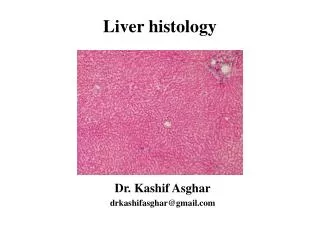
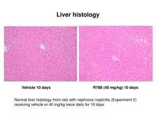
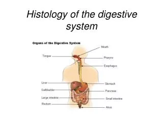
Histology of the Liver. The lobes of the liver are made up of lobules that contain hepatic cells ( liver cells or hepatocytes ), sinusoids , stellate reticuloendothelial ( Kupffer’s ) cells , and a central vein . Bile is secreted by hepatocytes.

E N D
Histology of the Liver • The lobes of the liver are made up of lobules that contain hepatic cells (liver cells or hepatocytes), sinusoids, stellate reticuloendothelial (Kupffer’s) cells, and a central vein. • Bile is secreted by hepatocytes. • Bile passes into bile canaliculi to bile ducts to the right and left hepatic ducts which unite to form the common hepatic duct. • Common hepatic duct joins the cystic duct to form the common bile duct which enters the hepatopancreatic ampulla.
Histology of the Gallbladder • Simple columnar epithelium • No submucosa • Three layers of smooth muscle • Serosa or visceral peritoneum
Application • Jaundice is a yellowish coloration of the sclera, skin, and mucous membranes due to a buildup of bilirubin. The main categories of jaundice are prehepatic, hepatic, and enterohepatic.
Blood Supply • The liver receives a double supply of blood from the hepatic artery and the hepatic portal vein. All blood eventually leaves the liver via the hepatic vein.
Bile - Overview • Hepatic cells (hepatocytes) produce bile that is transported by a duct system to the gallbladder for concentration and temporary storage. • Bile is partially an excretory product (containing components of worn-out red blood cells) and partially a digestive secretion. • Bile’s contribution to digestion is the emulsification of triglycerides. • The fusion of individual crystals of cholesterol is the beginning of 95% of all gallstones. Gallstones can cause obstruction to the outflow of bile in any portion of the duct system. Treatment of gallstones consists of using gallstone-dissolving drugs, lithotripsy, or surgery.
The liver also functions in carbohydrate, lipid, and protein metabolism; removal of drugs and hormones from the blood; excretion of bilirubin; synthesis of bile salts; storage of vitamins and minerals; phagocytosis; and activation of vitamin D. • In a liver biopsy a sample of living liver tissue is removed to diagnose a number of disorders.
Pathway of Bile Secretion • Bile capillaries • Hepatic ducts connect to form common hepatic duct • Cystic duct from gallbladder & common hepatic duct join to form common bile duct • Common bile duct & pancreatic duct empty into duodenum
Bile Production • One quart of bile/day is secreted by the liver • yellow-green in color & pH 7.6 to 8.6 • Components • water & cholesterol • bile salts = Na & K salts of bile acids • bile pigments (bilirubin) from hemoglobin molecule • globin = a reuseable protein • heme = broken down into iron and bilirubin
Liver Functions--Carbohydrate Metabolism • Turn proteins into glucose • Turn triglycerides into glucose • Turn excess glucose into glycogen & store in the liver • Turn glycogen back into glucose as needed
Liver Functions --Lipid Metabolism • Synthesize cholesterol • Synthesize lipoproteins----HDL and LDL(used to transport fatty acids in bloodstream) • Stores some fat • Breaks down some fatty acids
Liver Functions--Protein Metabolism • Deamination = removes NH2 (amine group) from amino acids so can use what is left as energy source • Converts resulting toxic ammonia (NH3) into urea for excretion by the kidney • Synthesizes plasma proteins utilized in the clotting mechanism and immune system • Convert one amino acid into another
Other Liver Functions • Detoxifies the blood by removing or altering drugs & hormones(thyroid & estrogen) • Removes the waste product--bilirubin • Releases bile salts help digestion by emulsification • Stores fat soluble vitamins-----A, B12, D, E, K • Stores iron and copper • Phagocytizes worn out blood cells & bacteria • Activates vitamin D (the skin can also do this with 1 hr of sunlight a week)
Summary of Digestive Hormones • Gastrin • stomach, gastric & ileocecal sphincters • Gastric inhibitory peptide--GIP • stomach & pancreas • Secretin • pancreas, liver & stomach • Cholecystokinin--CCK • pancreas, gallbladder, sphincter of Oddi, & stomach
Anatomy of the Small Intestine • 20 feet long----1 inch in diameter • Large surface area for majority of absorption • 3 parts • duodenum---10 inches • jejunum---8 feet • ileum---12 feet • ends at ileocecal valve
Mechanical Digestion in the Small Intestine • Segmentation, the major movement of the small intestine, is a localized contraction in areas containing food. • Peristalsis propels the chyme onward through the intestinal tract.
Mechanical Digestion in the Small Intestine • Weak peristalsis in comparison to the stomach---chyme remains for 3 to 5 hours • Segmentation---local mixing of chyme with intestinal juices---sloshing back & forth
Review: Digestion of Carbohydrates • Mouth---salivary amylase • Esophagus & stomach---nothing happens • Duodenum----pancreatic amylase • Brush border enzymes (maltase, sucrase & lactose) act on disaccharides • produces monosaccharides--fructose, glucose & galactose • lactose intolerance (no enzyme; bacteria ferment sugar)--gas & diarrhea
Review: Digestion of Proteins • Stomach • HCl denatures or unfolds proteins • pepsin turns proteins into peptides • Pancreas • digestive enzymes---split peptide bonds between different amino acids • brush border enzymes-----aminopeptidase or dipeptidase • enzymes break peptide bonds that attach terminal amino acids to carboxyl ends of peptides (carboxypeptidases) • enzymes break peptide bonds that attach terminal amino acids to amino ends of peptides (aminopeptidases) • enzymes split dipeptides to amino acids (dipeptidase)
Review: Digestion of Lipids • Mouth----lingual lipase • Most lipid digestion, in an adult, occurs in the small intestine. • emulsification by bile of globules of triglycerides • pancreatic lipase---splits triglycerides into fatty acids & monoglycerides • no enzymes in brush border
Digestion of Nucleic Acids • Nucleic acids are broken down into nucleotides for absorption. • Pancreatic juice contains 2 nucleases • ribonuclease which digests RNA • deoxyribonuclease which digests DNA • Nucleotides produced are further digested by brush border enzymes (nucleosidease and phosphatase) • pentose, phosphate & nitrogenous bases • Absorbed by active transport
Regulation of Secretion & Motility • Enteric reflexes that respond to presence of chyme • increase intestinal motility • VIP (vasoactive intestinal polypeptide) stimulates the production of intestinal juice • segmentation depends on distention which sends impulses to the enteric plexus & CNS • distention produces more vigorous peristalsis • 10 cm per second • Sympathetic impulses decrease motility
Regulation of Secretion & Motility • Enteric reflexes that respond to presence of chyme • increase intestinal motility • VIP (vasoactive intestinal polypeptide) stimulates the production of intestinal juice • segmentation depends on distention which sends impulses to the enteric plexus & CNS • distention produces more vigorous peristalsis • 10 cm per second • Sympathetic impulses decrease motility
Absorption of Monosaccharides • Essentially all carbohydrates are absorbed as monosaccharides. • They are absorbed into blood capillaries • Absorption of Amino Acids, Dipeptides, and Tripeptides • Most proteins are absorbed as amino acids by active transport processes. • They are absorbed into the blood capillaries in the villus
Anatomy of Large Intestine • 5 feet long by 2½ inches in diameter • Ascending & descending colon are retroperitoneal • Cecum & appendix • Rectum = last 8 inches of GI tract anterior to the sacrum & coccyx • Anal canal = last 1 inch of GI tract • internal sphincter----smooth muscle & involuntary • external sphincter----skeletal muscle & voluntary control
Appendicitis • Inflammation of the appendix due to blockage of the lumen by chyme, foreign body, carcinoma, stenosis, or kinking • Symptoms • high fever, elevated WBC count, neutrophil count above 75% • referred pain, anorexia, nausea and vomiting • pain localizes in right lower quadrant • Infection may progress to gangrene and perforation within 24 to 36 hours
Mechanical Digestion in Large Intestine • Mechanical movements of the large intestine include haustral churning, peristalsis, and mass peristalsis. • Peristaltic waves (3 to 12 contractions/minute) • haustral churning----relaxed pouches are filled from below by muscular contractions (elevator) • gastroilial reflex = when stomach is full, gastrin hormone relaxes ileocecal sphincter so small intestine will empty and make room • gastrocolic reflex = when stomach fills, a strong peristaltic wave moves contents of transverse colon into rectum
Absorption & Feces Formation in the Large Intestine • Some electrolytes---Na+ and Cl- • After 3 to 10 hours, 90% of H2O has been removed from chyme • Feces are semisolid by time reaches transverse colon • Feces = dead epithelial cells, undigested food such as cellulose, bacteria (live & dead)
Absorption and Feces Formation in the Large Intestine • The large intestine absorbs water, electrolytes, and some vitamins. • Feces consist of water, inorganic salts, sloughed-off epithelial cells, bacteria, products of bacterial decomposition, and undigested parts of food. • Although most water absorption occurs in the small intestine, the large intestine absorbs enough to make it an important organ in maintaining the body’s water balance.
Defecation Reflex • The elimination of feces from the rectum is called defecation. • Defecation is a reflex action aided by voluntary contractions of the diaphragm and abdominal muscles. The external anal sphincter can be voluntarily controlled (except in infants) to allow or postpone defecation.
Defecation • Gastrocolic reflex moves feces into rectum • Stretch receptors signal sacral spinal cord • Parasympathetic nerves contract muscles of rectum & relax internal anal sphincter • External sphincter is voluntarily controlled
Defecation Problems • Diarrhea = chyme passes too quickly through intestine • H20 not reabsorbed • Constipation--decreased intestinal motility • too much water is reabsorbed • remedy = fiber, exercise and water
Applications • Dietary fiber may be classified as insoluble (does not dissolve in water) and soluble (dissolves in water). • Both types affect the speed of food passage through the GI tract • Insoluble fiber • woody parts of plants (wheat bran, veggie skins) • may help protect against colon cancer • Soluble fiber • gel-like consistency = beans, oats, citrus white parts, apples • lowers blood cholesterol by preventing reabsorption of bile salts so liver has to use cholesterol to make more • Colonoscoy is the visual examination of the lining of the colon using an elongated, flexible, fiberoptic endoscope. • Occult blood test is to screen for colorectal cancer.
Regulation of Gastric Secretion and Motility • Cephalic phase • Gastric phase • Intestinal phase
Regulation of Gastric Emptying - Review • Release of chyme is regulated by neural and hormonal reflexes • Distention & stomach contents increase secretion of gastrin hormone & vagal nerve impulses • stimulate contraction of esophageal sphincter and stomach and relaxation of pyloric sphincter • Enterogastric reflex regulates amount released into intestines • distension of duodenum & contents of chyme • sensory impulses sent to the medulla inhibit parasympathetic stimulation of the stomach but increase secretion of cholecystokinin and stimulate sympathetic impulses • inhibition of gastric emptying
Vomiting (emesis) • Forceful expulsion of contents of stomach & duodenum through the mouth • Cause • irritation or distension of stomach • unpleasant sights, general anesthesia, dizziness & certain drugs • Sensory input from medulla cause stomach contraction & complete sphincter relaxation • Contents of stomach squeezed between abdominal muscles and diaphragm and forced through open mouth • Serious because loss of acidic gastric juice can lead to alkalosis
Aging and the Digestive System • Changes that occur • decreased secretory mechanisms • decreased motility • loss of strength & tone of muscular tissue • changes in neurosensory feedback • diminished response to pain & internal stimuli • Symptoms • sores, loss of taste, peridontal disease, difficulty swallowing, hernia, gastritis, ulcers, malabsorption, jaundice, cirrhosis, pancreatitis, hemorrhoids and constipation • Cancer of the colon or rectum is common