Download
1 / 22
270 likes | 963 Vues
HISTOLOGY OF THE BLOOD VESSELS. By the end of this lecture, the student should be able to identify and describe the microscopic structure of the wall of the blood vessels including: a. Elastic arteries. b. Muscular (medium-sized) arteries. c. Medium-sized veins.

E N D
HISTOLOGY OF THE BLOOD VESSELS By the end of this lecture, the student should be able to identify and describe the microscopic structure of the wall of the blood vessels including: a. Elastic arteries. b. Muscular (medium-sized) arteries. c. Medium-sized veins. d. Blood capillaries.
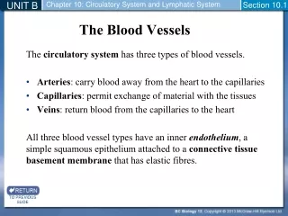
Blood vessels: • Arteries: • Elastic artery. • Muscular (distributing) (medium-sized) artery. • Arterioles. • Blood capillaries. • Veins: • Venules. • Small veins. • Medium-sized veins. • Large veins.
General Structure of Blood Vessels • The wall of blood vessel is formed of three concentric layers: • Tunica intima (interna) • Tunica media • Tunica adventitia (externa)
Tunica Intima • Is the innermost layer • Composed of: • Endothelial cells: Simple squamous epithelium • Subendothelial layer: loose C.T. • Internal elastic lamina: fenestrated elastic sheet.
Tunica Media • Intermediate layer • Composed of: • Smooth muscles. • Elastic fibers. • Type III collagen (reticular fibers). • Type I collagen. NB: Large muscular arteries have external elastic lamina, separating the tunica media from the tunica adventitia
Tunica Adventitia • Outermost layer • Composed of connective tissue containing Vasavasorum: They are small arterioles in tunica adventitia and the outer part of tunica media. They are more prevalent in the walls of veins than arteries – why? Venous blood contains less oxygen and nutrients than arterial blood.
ELASTIC ARTERIES • Examples: aorta, common carotid a., subclavian a., common iliac a, pulmonary Trunk. • Microscopic structure: • T. Intima: *Endothelium. *Subendothelial C.T. *Internal elastic lamina: (not prominent) (indistinct)
ELASTIC ARTERIES (Cont.) • T. Media: it consists of: • Fenestrated elastic membranes (sheets) (lamellae): It is the main component of T.M. B. In between, there are: • Smooth muscle cells. 2. Collagen fibers (type I collagen). 3. Reticular fibers (type III collagen). 4. Elastic fibers.
ELASTIC ARTERIES (Cont.) • T. Adventitia: • Much thinner than T.M. • It is composed of loose C.T. • Contains vasa vasorum → send branches to the outer part of T.M.
MUSCULAR ARTERIES(Medium-sized artery) • Examples: brachial, ulnar, renal. • Microscopic structure: • T. Intima.: • Endothelium. • Subendothelial C.T. layer. • Internal elastic lamina: • Is prominent. • Displays an undulating surface.
MUSCUALR ARTERIES (Cont.) • T. Media:(Thicker than T. Adventitia or similar in thickness). Components: • Smooth muscle cells (SMCs): are the predominant component. B. In between there are: • Elastic fibers. • Type III collagen fibers. • Type I collagen fibers. • External elastic lamina: may be identifiable. 3. T. Adventitia.: loose C.T.
MEDIUM-SIZED VEIN • Thickness of the wall: thinner than the accompanying artery. T. Intima: *usually forms valves. *no internal elastic lamina • T. Media: • Thinner than T. Adventitia • Consists of: • Fewer SMCs. • Types I & III Collagen fibers. • T. Adventitia: thicker than T. Media
VALVES OF VEINS • Valve of a vein is composed of 2 leaflets • Each leaflet has a thin fold of the T. Intima. • Components: • Endothelium • Core of C.T.
BLOOD CAPILLARIES • Diameter: usually 8-10 µm. • Microscopic structure: • Single layer of squamous endothelial cells. • Basal lamina: surrounds the external surface of the endothelial cells. • Pericytes: • Have processes. • Share the basal lamina of the endothelial cells.
BLOOD CAPILLARIES Types: 1- Continuous blood capillaries 2- Fenestrated blood capillaries a- with diaphragms b- without diaphragms 3- Sinusoidal blood capillaries
Continuous Blood Capillaries • Microscopic structure: • No pores or fenestrae in their walls. • Distribution: • In muscles, nervous T., C.T.
Fenestrated Blood Capillaries with Diaphragms • Microscopic structure: • The walls of their endothelial cells have pores (fenestrae). • These pores are covered by diaphragm. • Distribution: • In intestine, pancreas and endocrine glands.
Fenestrated Blood Capillarieswithout Diaphragms • Microscopic structure: • The walls of their endothelial cells have pores (fenestrae). • These pores are NOT covered By diaphragm. • Distribution: In renal glomerulus.
FENESTRATED CAPILLARYWITH DIAPHRAGMS FENESTRATED CAPILLARYWITHOUT DIAPHRAGMS Lumen of glomerular Blood capillary Endothelium Podocyte
SINUSOIDAL CAPILLARIES • Diameter: irregular (30-40 µm). • Microscopic features: • Their endothelial cells have fenestrae without diaphragms. • They possess discontinuous endothelial cells. • They possess discontinuous basal lamina. • Macrophages may be located in or along the outside of the endothelial wall. • Distribution: Red bone marrow, liver, spleen and certain endocrine glands.