Download
1 / 21
220 likes | 313 Vues
Explore the structure and function of arteries, capillaries, and veins in the circulatory system. Learn how blood flows and exchanges nutrients. Discover the role of lymphatic vessels in maintaining fluid balance.

E N D
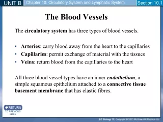
UNIT B Chapter 10: Circulatory System and Lymphatic System Section 10.1 The circulatory system has three types of blood vessels. Arteries: carry blood away from the heart to the capillaries Capillaries: permit exchange of material with the tissues Veins: return blood from the capillaries to the heart All three blood vessel types have an inner endothelium, a simple squamous epithelium attached to a connective tissue basement membrane that has elastic fibres. The Blood Vessels TO PREVIOUS SLIDE
UNIT B Chapter 10: Circulatory System and Lymphatic System Section 10.1 The largest artery in the body is the aorta, which carries O2-rich blood from the heart to other parts of the body. The arterial wall has three layers. Inner layer: endothelium Middle layer: smooth muscle that contracts and relaxes to regulate blood flow and pressure Outer layer: fibrous connective tissue The Arteries Figure 10.1 Blood vessels. The walls of arteries and veins have three layers. The inner layer is composed largely of endothelium, with a basement membrane that has elastic fibres; the middle layer is smooth muscle tissue; the outer layer is connective tissue (largely collagen fibres). a. Arteries have a thicker wall than veins because they have a larger middle layer than veins. TO PREVIOUS SLIDE
UNIT B Chapter 10: Circulatory System and Lymphatic System Section 10.1 Arterioles are small arteries that branch off from an artery. Arterioles have three layers. Inner layer: endothelium Middle layer: some elastic tissue but mostly smooth muscle Smooth muscle contracts: blood vessel constricts, resulting in higher blood pressure Smooth muscle relaxes: blood vessel relaxes, resulting in lower blood pressure Outer layer: fibrous connective tissue TO PREVIOUS SLIDE
UNIT B Chapter 10: Circulatory System and Lymphatic System Section 10.1 Veins and venules (small veins) take blood from the capillary beds to the heart. Veins and venules have the same three layers as arteries, but there is less smooth muscle and connective tissue Veins have valves that prevent backflow The Veins TO PREVIOUS SLIDE
UNIT B Chapter 10: Circulatory System and Lymphatic System Section 10.1 Veins act as a blood reservoir Since their walls are thinner, they can expand to a greater extent About 70% of blood is in the veins The largest veins in the body are the venae cavae (superior vena cava, inferior vena cava), which deliver O2-poor blood to the heart TO PREVIOUS SLIDE
UNIT B Chapter 10: Circulatory System and Lymphatic System Section 10.1 Capillaries are narrow blood vessels that join arterioles to venules. Composed of a single layer of epithelium with a basement membrane Form vast networks (capillary beds) throughout the body The Capillaries TO PREVIOUS SLIDE
UNIT B Chapter 10: Circulatory System and Lymphatic System Section 10.1 Only certain capillary beds are open at any given time. After eating, capillary beds that serve the digestive system are open, and those that serve the muscles are mostly closed Sphincter muscles relax to open the bed and allow blood flow Sphincter muscles contract to close the bed and prevent blood flow When the bed is closed, blood flows through anastomoses(arteriovenous shunts) directly from arterioles to venules, bypassing the bed TO PREVIOUS SLIDE
UNIT B Chapter 10: Circulatory System and Lymphatic System Section 10.1 Figure 10.2 Anatomy of a capillary bed. A capillary bed forms a maze of capillary vessels that lies between an arteriole and a venule. When precapillary sphincter muscles are relaxed, the capillary bed is open, and blood flows through the capillaries. When sphincter muscles are contracted, blood flows through a shunt (anastomosis) that carries blood directly from an arteriole to a venule. As blood passes through a capillary in the tissues, it gives up its oxygen. Therefore, blood goes from being O2-rich in the arteriole (red colour) to being O2-poor in the vein (blue colour). TO PREVIOUS SLIDE
CAPILLARY FLOW • Two forces control movement of fluid through capillary walls: • Osmotic pressure: draw of fluid to the dissolved solutes that can’t cross semipermeable membrane • Tends to cause water to move from tissue fluid to blood • Created by salts and plasma proteinds • Hydrostatic Pressure (Blood Pressure): tends to cause water to move in the opposite direction.
UNIT B Chapter 10: Circulatory System and Lymphatic System Section 10.2 . TO PREVIOUS SLIDE
UNIT B Chapter 10: Circulatory System and Lymphatic System Section 10.1 Exchange of substances takes place across the thin walls of the capillaries. Oxygen and nutrients diffuse out of the capillary and into the tissue fluid that surrounds cells Wastes (carbon dioxide) diffuse into the capillary Some water leaves the capillaries, and excess is picked up by lymphatic vessels TO PREVIOUS SLIDE
UNIT B Chapter 10: Circulatory System and Lymphatic System Section 10.2 Fluid in the blood is called plasma. When blood reaches a capillary, the movement of fluid in the blood through the capillary wall is controlled by: Osmotic pressure (causes water to move from the tissue fluid to the blood) Blood pressure (causes water to move from blood to tissue fluid) Capillary Exchange TO PREVIOUS SLIDE
UNIT B Chapter 10: Circulatory System and Lymphatic System Section 10.2 Arterial End of Capillary Blood pressure (hydrostatic pressure) is higher than osmotic pressure of blood Water exits capillary Midway Along the Capillary Blood pressure and osmotic pressure cancel each other out No net movement of water Solutes diffuse according to concentration gradient Nutrients and oxygen diffuse out of the capillary; wastes and carbon dioxide diffuse into the capillaries Small substances leaving capillaries contribute to tissuefluid TO PREVIOUS SLIDE
UNIT B Chapter 10: Circulatory System and Lymphatic System Section 10.2 Venous End of Capillary Osmotic pressure is greater than blood pressure Water moves into capillary Excess tissue fluid is collected by lymphaticcapillaries, where it becomes lymph Figure 10.10 Lymphatic capillaries. A lymphatic capillary bed (shown here in green) lies near a blood capillary bed. When lymphatic capillaries take up excess tissue fluid, it becomes lymph. TO PREVIOUS SLIDE
UNIT B Chapter 10: Circulatory System and Lymphatic System Section 10.1 Describe how blood flow is controlled in each of the three major types of blood vessels. List several specific substances that diffuse across capillary walls. Check Your Progress TO PREVIOUS SLIDE
UNIT B Chapter 10: Circulatory System and Lymphatic System Section 10.1 TO PREVIOUS SLIDE