Download
1 / 50
510 likes | 1.28k Vues
THE CHROMOSOMAL BASIS OF INHERITANCE. Unit 3- Chapter 15. A. A. A. A. B. B. B. B. B. B. A. A. A. A. B. B. Non-linked vs. Linked Genes. If the genes in question are on different chromosome pairs, they are considered non-linked.

E N D
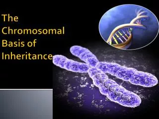
THE CHROMOSOMAL BASIS OF INHERITANCE Unit 3- Chapter 15
A A A A B B B B B B A A A A B B Non-linked vs. Linked Genes • If the genes in question are on different chromosome pairs, they are considered non-linked. • This PGC will give rise to only one kind of gamete with respect to the genes A and B., because it is homozygous for both traits. Primordial Germ Cells Gametes • If the genes in question are on the same chromosome pair, they are considered linked. • This PGC will also give rise to only one kind of gamete, with respect to the genes A and B, because it is homozygous for both traits.
A a A a b B B b b B B b a A a a A A B b Non-linked vs. Linked Genes Primordial Germ Cell • In this case, the PGC is heterozygous for both traits, and the genes are non-linked, so after meiosis, it can produce 4 different types of gametes. • In this PGC however, the genes in question are lined. So even though the PGC is heterozygous for both traits, after meiosis, it can only produce 2 different types of gametes.
a A A A a a b B B b B B b b a A What is the outcome of this cross? P generation X Gametes F1 generation Phenotype = 100% dominant Genotype = 100% heterozygous
A A a a B b B b B b B b B B b b a A a a A A A a The F2 Generation in non-linked genes P2 generation X Gametes F2 generation The phenotypes of the F2 generation in a dihybrid cross of non-linked genes always results in a 9:3:3:1 ratio
A a A A a a B b b b B B b a A What is the outcome of this cross? P generation X Gametes B F1 generation Phenotype = 100% dominant Genotype = 100% heterozygous
A A a a b b B B B B b A a a A b The F2 Generation in linked genes P2 generation X Gametes F2 generation Genotype = 1:2:1 Phenotype = 3:1 dominant
Recombination in non-linked genes – involves independent assortment of chromosomes In nonlinked genes, the following test cross: YyRr X yyrr Would yield a phenotypic ratio of 1:1:1:1
Recombination in linked genes – involves crossing over Thomas Morgan, noticed something strange when working with linked genes.
Crossing over created more types of gametes than what Morgan expected
Linkage maps Recombination frequencies can be used to create linkage maps, which indicate the distance between various genes on the same chromosome. The distance is measured in “centimorgans” or “map units”. The recombination frequency cannot exceed 50%, because after that, one cannot tell linked from nonlinked genes.
Linked Genes • Genes located on the same chromosomes • They tend to be inherited together - are not sorted independently like unlinked genes usually are • However, they can be “shuffled” through crossing over or recombination • The farther apart the genes are from each other in a chromosome, the greater the likelihood that they will be unlinked as a result of crossing-over. In this example, the "A" and "E" genes have the highest likelihood of being unlinked because they are at opposite ends of their chromosome.
SEX-LINKED inheritance This involves genes which are found only on the X chromosome. The pattern of inheritance differs from somatic genes, because males have only one X chromosome.
Sex-Linked Inheritance, cont’d. • In humans and fruit flies, • females have two X chromosomes • males have one X and one Y (HEMIZYGOUS – only one copy of a gene on the XY chromosomes) • females get an X from the mother and another X from the father • males get an X from the mother and a Y from the father • In humans, the Y chromosome is relatively small and contains only a few genes, among them the important "SRY" gene, which contains the instructions for turning on genes on other chromosomes to activate the signals for making male hormones and male anatomical peculiarities. The SRY gene and some other "Y" chromosome genes are not present on the X chromosome • In humans and also fruit flies, the X chromosome contains many genes which are not present on the Y chromosome. For these genes males have only one allele.
Hemophilia Perhaps the most well-known disease related to plasma is hemophilia. An inherited change in one of the clotting proteins (called factor VIII) leaves it dysfunctional. This single change disrupts the entire sequence of chemical reactions necessary for clotting. As a result, people with hemophilia can suffer severe swelling, bruising and bleeding from simple day to day events that the rest of the population take for granted. Hemophilia The child shown above has a severe swelling on the forehead from a bump to the head. This was caused by a failure of his clotting system to stop the bleeding from a bruise. Hemophilia can be controlled with the infusion of factor VIII collected from donated blood or plasma.
X-Inactivation n Females • One X chromosome condenses into a compact object visible as a dark spot against the inner nuclear envelope -this is called the Barr Body • This happens during early embryonic development • Therefore, males and females have in reality, only one active X chromosome
Barr Bodies Mosaicism
Hemophilia in Females Hemophilia B is an X-linked bleeding disorder resulting from factor IX (F.IX) deficiency, caused by a wide range of mutations on the F.IX gene. Hemophilia B in girls is extremely rare and results from different mechanisms, the most common of which is skewed inactivation of the normal X chromosome in heterozygous girls. In some cases, the inactivation process does not seem to be random and occurs by either selection in favor of the activity of an X chromosome involved in a balanced X:autosome translocation or as a result of genetic differences affecting the X chromosome inactivation itself. More rarely, the phenotypic expression of the disease can be related to compound heterozygosity for hemophilic mutations or Turner syndrome.
NON-DISJUNCTION NORMAL DISJUNCTION NON-DISJUNCTION Results of non-disjunction: Aneuploidy - Trisomy, Monosomy, Polyploidy
Down Syndrome, Trisomy 21 Down Syndrome can also be caused by the translocation of chromosomes (most often chromosome 14 or 21). Frequency of Down Syndrome increases with the mother’s age.
Turner Syndrome • The most common characteristics of Turner syndrome include short stature and lack of ovarian development. A number of other physical features, such as webbed neck, arms that turn out slightly at the elbow, and a low hairline in the back of the head are sometimes seen in Turner syndrome patients. Individuals with Turner syndrome are also prone to cardiovascular problems, kidney and thyroid problems, skeletal disorders such as scoliosis (curvature of the spine) or dislocated hips, and hearing and ear disturbances.
Cri du chat • Major identifying characteristics • Monotone, weak, cat-like cry • Small head (microcephally) • High palate • Round face • Small receding chin (micrognathia) • Widely spaced eyes (hypertelorism) • Low set ears • Low broad nasal ridge • Folds of skin over the upper eyelid (epicanthic folds) • Distinctive creases on the palms of the hands
Chromosome 5 Cri Du Chat Syndrome results from the loss or deletion of a significant portion of the genetic material from the short arm of one of the pair of number five chromosomes. Cri Du Chat Syndrome is also known as 5P Minus syndrome, Le Jeune's syndrome and Cat's-cry syndrome. It is possible to detect Cri du Chat Syndrome with amniocentesis or CVS (Chorionic Villus Sampling) in the first trimester of pregnancy. An ultrasound may lead the doctor to suspect a disorder of this type and carry out further investigations but it is not possible to diagnose it solely by this means. The critical region of the chromosome containing genes which are responsible for the main features of the syndrome appears to be located in band 5p15.2. The gene causing the cry has been located in band 15.3. This would explain why some babies with other features of the syndrome do not have the characteristic cry and some babies have the cry but not the other characteristics.
Genomic Imprinting • Genes remember? • Alleles that come from mother (ovum) may have a different effect on the individual than alleles that come from father (sperm) • This pattern continues for all future generations. • Example: Prader-Willi / Angelman Prader-Willi- mutation in gene on Ch.15: If inherited from father, child gets the disease. The mother’s genes in this area are “turned off” through DNA methylation • If inherited from mother, child gets Angelman’s syndrome. Father’s gene is methylated and inactive.
Prader-Willi Syndrome • characterizedby • severe hypotonia and feeding difficulties in early infancy, followed in later infancy or early childhood by excessive eating and gradual development of morbid obesity. • Motor and language development are delayed. • All individuals have some degree of cognitive impairment. • A distinctive behavior (with temper tantrums, stubbornness, manipulative behavior, and obsessive-compulsive characteristics) is common. • Hypogonadism is present in both males and females and manifests as genital hypoplasia, incomplete pubertal development, and, in most, infertility. • Short stature is common; characteristic facial features, strabismus, and scoliosis are often present, and • non-insulin-dependent diabetes mellitus often occurs in obese individuals.
Angelman Syndrome • characterized by • Developmental delay, functionally severe • Speech impairment, none or minimal use of words; receptive and non-verbal communication skills higher than verbal ones • Movement or balance disorder, usually ataxia of gait and/or tremulous movement of limbs • Behavioral uniqueness: any combination of frequent laughter/smiling; apparent happy demeanor; easily excitable personality, often with hand flapping movements; hypermotoric behavior; short attention span
How does the cell express only certain genes? • The cell uses 2 techniques in order to have certain genes (or alleles) expressed and others silenced. • Methylation of cysteines in gene (DNA segment) • Acetylation of histones in the region of the gene
DNA methylation • Enzymes (DNA methyl transferases)add a methyl group (--CH3) to the number 5 carbon of the cytosine pyrimidine ring. • This causes that segment of DNA to become compact – it is inaccessible to enzymes and cannot be transcribed into mRNA. • So the gene is silent. This compact DNA stains dark and is called heterochromatin.
Histone Acetylation • Acetyl groups (COCH3) are added by enzymes to the tails of histone proteins • This prevents the DNA from “hugging” the histones, making that segment of DNA more accessible to transcription enzymes. • So this gene is expressed • Opposite effect of DNA methylation
"CpG" stands for cytosine and guanine separated by a phosphate • (C-phosphate-G-), which links the two nucleosides together in DNA. • The "CpG" notation is used to distinguish a cytosine followed by guanine, from a cytosine base-paired to a guanine.
Inheritance of mDNA* and pDNA* • Mitochondria are always inherited from the mother (In animals and plants) • Daughters will pass them on to their progeny • Sons will not pass them on to their offspring • Plastids are always inherited from the plant ova (egg) and not from the pollen *mDNA=mitochondrial DNA *pDNA=plastid DNA
mDNA mutations • mDNA contains genes for proteins used in the electron transport chain • Defects in these genes will cause problems with ATP production • Less ATP = decreased muscle and neuron function, because muscle cells and neurons have high energy (ATP) requirement.