Protista
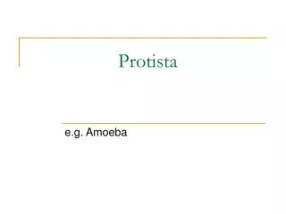
Protista. The “Leftovers” Kingdom. General Characteristics. All contain a nucleus in each cell Most live in watery environments Most are single-celled organisms


Protista
E N D
Presentation Transcript
Protista The “Leftovers” Kingdom
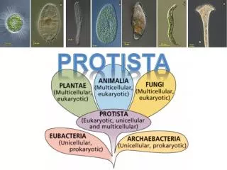
General Characteristics • All contain a nucleus in each cell • Most live in watery environments • Most are single-celled organisms • Most live as individual cells but many live as a colony ( a number of relatively independent cells of the same species that are attached to one another)
General Characteristics • Evolved ~ 1.5 billion years ago • Some are autotrophs, some are heterotrophs and some can be either • Three general categories • Animal-like • Plant-like • Fungus-like
Also called protozoa (= first animals) Characteristics of: Nucleus No cell wall Heterotrophs Most can move 4 types: Sarcodines Ciliates Zooflagellates Sporozoans Animal-like Protists
Sarcodines • Characterized by extensions of the cell membrane and cytoplasm known as pseudopods (pseudopod = false foot) • Pseudopods are used to capture and engulf particles of food and to move from one place to another
Sarcodines • Feed by the process of phagocytosis Illustration modified from public domain image http://commons.wikimedia.org/wiki/File:Amoeba_fagocitose.jpg
Sarcodines – with Shells • Many sarcodines have shells (also called tests) • The shells must have openings that allow the pseudopods to extend out • These shells are part of limestone, marble and chalk • Two examples of shelled sarcodines are foraminiferans and radiolarians
Sarcodines -Foraminiferans Foraminifera "Star sand" Hatoma Island – Japan Photo by Psammophilehttp://en.wikipedia.org/wiki/File:2085f_Japon_Hatoma.jpg • Their shells are made of calcium carbonate or organic materials. FYI: *Commonly found on the Abyssal plains, at depths of up to 6.6 miles. ** Among the largest known unicellular organisms. Although they are unicellular, these shelled ameboids can be seen without a microscope. They typically are less than 1mm in diameter, but some found on the deep ocean floor*, may reach 20 cm in diameter**.
Sarcodines – Foraminiferans Tests of foraminifera extracted sand from the beach of Ngapali (Myanmar) Photo by: Psammophile http://en.wikipedia.org/wiki/File:Foraminif%C3%A8res_de_Ngapali.jpg SEM micrographs of four benthic foraminiferans from the USGS (Public Domain: http://en.wikipedia.org/wiki/File:Benthic_foraminifera.jpg ) FYI: Benthic means bottom dweller.
Sarcodines – Foraminiferans Live Ammonia tepida benthic foraminiferan collected from San Francisco Bay. Phase-contrast photomicrograph by Scott Fay, UC Berkeley, 2005. http://en.wikipedia.org/wiki/File:Ammonia_tepida.jpg Ammonia beccarii , benthic forams collected in 2011 on the edge of the Belgian part of the North Sea. Photo by: Hans Hillewaert http://en.wikipedia.org/wiki/File:Ammonia_beccarii.jpg
Sarcodines - Radiolarians • Radiolarians are zooplankton that have a mineral skeleton containing silica • Their skeletons can be very elaborate. • They range in size from 0.1-0.2 mm in diameter. Illustration by Ernst Haeckel, 1904, Public Domain http://en.wikipedia.org/wiki/File:Haeckel_Spumellaria.jpg FYI: Plankton are organisms that float in the ocean.
Sarcodines - Radiolarians Various Radiolaria Photo by: Luis Fernández García http://commons.wikimedia.org/wiki/File:Radiolaria_varios.jpg REM(Reflection Electron Micrograph) of a Radiolarian Photo by Hannes Grobe/AWI http://commons.wikimedia.org/wiki/File:Radiolaria_hg.jpg Skeleton of a polycystine radiolarian Public Domain: NASA http://en.wikipedia.org/wiki/File:Radiolarian.png
Sarcodines – Amebas (or Amoeba) • Reproduce by binary fission (the same as bacteria) – one cell divides into two • Respond to light and certain chemicals by moving away • Because they live in fresh water they have a special structure, the contractile vacuole, that pumps excess water out of the cell. Water diffuses in by osmosis and must be actively pumped out or the cell would burst.
Sarcodines - Ameba • Cytoplasm • Nucleus • Contractile Vacuole • Cell Membrane • Food Vacuole • Pseudopods Racette
Sarcodines - Amebas Chaos carolinensis a species of giant ameba (up to 5mm) Photo by: Dr.Tsukii Yuuji http://pl.wikipedia.org/wiki/Plik:Chaos_carolinense.jpg Amoeba proteus Photo by Cymothoa exigua http://commons.wikimedia.org/wiki/File:Amoeba_proteus.jpg
Ameba Videos Ameba in motion https://www.youtube.com/watch?v=7pR7TNzJ_pA Ameba feeding https://www.youtube.com/watch?v=W6rnhiMxtKU https://www.youtube.com/watch?v=x1ErCyZCFw8
Ciliates • All have cilia – small hair-like projections on the outer surface of the cell • Cilia do three things • Act like tiny oars to help these organisms move • Sweep food particles towards themselves • Act as tiny sensors
Ciliates • Have two nuclei • Large nucleus (Macronucleus) controls cell functions • Small nucleus (Micronucleus) controls the process of conjugation • Conjugation – two ciliates (same species) temporarily join and exchange part of their DNA • Conjugation is followed by binary fission Ciliate undergoing binary fission Photo by TheAlphaWolf http://en.wikipedia.org/wiki/File:Unk.cilliate.jpg
Ciliates - Paramecium Paramecium caudatum Photo by Deuterostome http://en.wikipedia.org/wiki/File:Paramecium_caudatum_Ehrenberg,_1833.jpg Paramecia, illustrated by Otto Müller, 1773.
Ciliates - Paramecium • Pellicle – the cell membrane and underlying structures that give the organism its shape; without the pellicle a paramecium would look like a hairy ameba • Gullet – a funnel-like structure that captures food into food vacuoles • Oral Groove – guides food to the gullet • Anal Pore – empties wastes out of the cell
Ciliates - Paramecium • Have contractile vacuoles to pump out water; look a bit like a flower • Video: https://www.youtube.com/watch?v=mTXRcbjuYGU Part of a photo by Barfooz http://commons.wikimedia.org/wiki/File:Paramecium.jpg C.V.
Ciliates - Paramecium Illustration modified from Public Domain image http://commons.wikimedia.org/wiki/File:Paramecium_sp.jpg
Other Ciliates Blepharisma japonicum Photo by Frank Fox (www.mikro-foto.de) onhttp://en.wikipedia.org/wiki/Blepharisma Tetrahymena thermophila Photo Source: Ciliate Genome Sequence Reveals Unique Features of a Model Eukaryote. Robinson R, PLoS Biology Vol. 4/9/2006, e304. doi:10.1371/journal.pbio.0040304 (Creative Commons License) Vorticella Photo by Frank Fox (www.mikro-foto.de) at http://commons.wikimedia.org/wiki/File:Mikrofoto.de-Vorticella_7.jpg
Other Ciliates - Stentor A composite image of Stentor roeseli This work has been released into the public domain by its author, Protist Image Database http://en.wikipedia.org/wiki/File:Stentor_roeseli_composite_image.jpg
Zooflagellates • Flagellates are protists that move by means of a flagellum, a long, whip-like structure. They may be animal-like, plant-like or fungus-like. • Zooflagellates are animal-like flagellates.
Zooflagellates • Usually have 1-8 flagella, depending on the species • Many live inside the bodies of animals (they are symbiotic). • Some are mutualistic – for example those that live in the gut of the termite and digest wood • Many are parasitic – Examples include Typanosoma sp. (causes African Sleeping Sickness) and Giardia
Zooflagellates False color SEM of Trypanosoma brucei (found in the gut of the tsetse fly host) Photo by Zephyris http://commons.wikimedia.org/wiki/File:TrypanosomaBrucei_ProcyclicTrypomastigote_SEM.jpg Giardia Public Domain: Centers for Disease Control and Prevention http://en.wikipedia.org/wiki/File:Giardia_lamblia_SEM_8698_lores.jpg Trypanosoma evansi Photo by Alan R Walker http://commons.wikimedia.org/wiki/File:Trypanosoma-evansi.jpg
Sporozoans • All are parasites that feed on the cells and body fluids of their host animals • Many have complex life cycles with more than one host • Each forms a spore during some part of their life cycle that enables it to be passed from host to host
Sporozoans - Plasmodium • Several different species of Plasmodium cause the disease malaria. • They are transmitted only by Anopheles mosquitos. FYI: According to the WHO, there were an estimated 207 million cases of malaria in 2012 (uncertainty range: 135 – 287 million) and an estimated 627 000 deaths (uncertainty range: 473 000 – 789 000). Ninety percent of all malaria deaths occur in sub-Saharan Africa, 75% of whom were children under the age of five.
Mosquito Photo Credit: James Gathany, CDC http://en.wikipedia.org/wiki/File:Anopheles_albimanus_mosquito.jpg Plasmodium Life Cycle Each sporozoan divides to form many spores Sporozoans develop in the mosquito’s gut Spores move to the mosquito’s mouth Female Anopheles albimanus mosquito, transmits malaria in Central America Mosquito bites human injecting spores Mosquito bites human and drinks infected blood In Mosquito Host In Human Host Spores travel to the liver Red blood cells burst releasing sporozoans that then infect more RBCs Infect liver cells, multiply and burst out Sporozoans infect red blood cell and multiply Liver and Blood cell diagrams part of Public Domain image from NIH http://sw.wikipedia.org/wiki/Picha:MalariacycleBig.jpg