Download
1 / 69
690 likes | 708 Vues
Learn about esophageal motor disorders including Smooth Muscle Achalasia and Diffuse Esophageal Spasm, their clinical features, diagnosis using manometry and barium swallow, and available treatments.

E N D
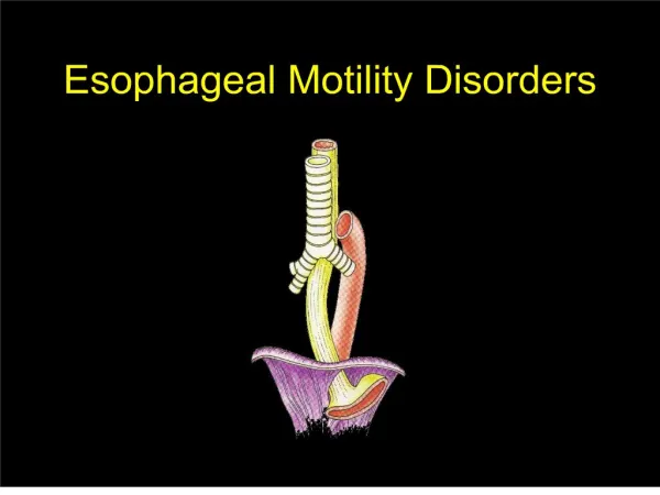
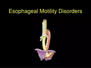
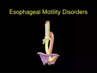
the 2 major functions of the esophagus: • transport of the food bolus from the mouth to the stomach • the prevention of retrograde flow of GI contents • the UES remains closed due to • the elastic properties of its wall • tonic contraction of the cricopharyngeus and inferior pharingeal constrictor muscles ( produced by continuous neural excitation of the lower motor neurons which innervate these muscles via end plates) • in contrast, the LES remains closed because of its intrinsec myogenic tone and a neural pathway, consisting of preganglionic parasympathetic fibers in the vagus nerve and postganglionic myenteric inhibitory neurons, causes its relaxation.
Pathophysiology • defective inervation of the smooth muscle portion of the esophageal body and LES • vigorous achalasia less severe neural damage than classic one ( shows a marked reduction in myenteric neurons)
Clinical features All ages, both sexes • Symptoms: • Dysphagia • early with both liquids and solids • worsened by emotional stress and hurried eating • Chest pain • Regurgitation and pulmonary aspiration (occurs because of retention of large volumes of saliva and ingested food in the esophagus) • The overall course is usually chronic with progressive dysphagia and weight loss over month to years
Manometry • the esophageal body shows elevated resting pressure • in response to swallows, primary peristaltic waves are replaced by simultaneous onset contractions. • these contractions may be of: • large amplitude and long duration (vigorous A) • poor amplitude (classic A) • administration of the cholinergic muscarinic agonist mecholyl causes a marked increase in baseline esophageal pressure and CCK paradoxically causes contraction of LES ( normally causes a fall in the sphincter pressure )
Manometric representation of normal peristalsis and achalasia. • Measurements are taken from multiple recording levels in the esophagus. • In this example of achalasia, • peristalsis and lower esophageal sphincter (LES) relaxation are absent and • LES pressure is elevated. • AA=aortic arch; S= swallow.
TREATMENT • MEDICAL TREATEMENT: • soft foods • sedatives • nitrates • anticholinergic drugs usually unsatisfactory • Calcium channel antagonists (NIFEDIPINE) • BALLON DILATATION –the best available therapy, effective in 85% of patients; complications: perforation, bleeding • HELLER’S EXTRAMUCOSAL MYOTOMY OF LES
COMPLICATIONS On the far left an intramural extravasation (arrow) after distal dilation for achalasia. In the middle an intramural extravasation (arrow) after complicated endoscopy. On the right a perforation after biopsy with extravasation of contrast material (arrow).
Definition: motor disorder of the esophageal smooth muscle with multiple spontaneous contractions and swallow induced contractions that are of simultaneous onset , large amplitude and repetitive occurrence. Variants of DES ( NUTCRACKER ESOPHAGUS) occur as a primary disease or in association with a variety of diseases, as well as emotional stress and aging: • Collagen vascular disease • Diabetic neuropathy • Reflux esophagitis • Irradiation esophagitis • Esophageal obstruction • Drugs ( cholinergic , anticholinergic)
Clinical features • Chest pain ± retrosternal , at rest, emotional stress • Dysphagia for solids and liquids • Radiation –to the back - sides of the chest - both arms - sides of the jaw • may last for a few seconds to several minutes • acute and severe ≠ myocardial ischemia (angina)
Diagnosis uncoordinated simultaneous contractions that produce curling or multiple ripples in the wall, sacculation and pseudodiveticula – “the CORK SCREW “E. LES opens normally BARIUM SWALLOW 74-year-old man with diffuse esophageal spasm who presented with dysphagia. Prone right anterior oblique view from single-contrast esophagram shows multiple nonperistaltic contractions (arrows) of moderate severity without classic corkscrew appearance. Lower esophageal sphincter opened normally.
Diagnosis reveals prolongued large amplitude and repetitive contractions of simultaneous onset MANOMETRY Manometric representation of normal peristalsis and diffuse esophageal spasm. Measurements are taken from multiple recording levels in esophagus. Normal peristalsis is present in upper esophagus, but it is replaced by simultaneous, repetitive contractions below aortic arch (AA). Normal lower esophageal sphincter (LES) relaxation is seen. S, swallow.
MANOMETRY Esophageal manometry tracing demonstrates nutcracker esophagus. Note the excessive amplitude of the contractions. This is normal esophageal manometry tracing with normal amplitude of the contractions. The contractions are coordinated because the contractions in the proximal esophagus (top of image) occur before the contractions further distal in the esophagus. Esophageal manometry tracing demonstrates diffuse esophageal spasm. Note the multiple uncoordinated contractions in the third tracing from the distal esophagus
Diagnosis • Cold swallows and solid boluses induce chest pain and motor abnormalities • Provocative pharmacologic tests LIMITED • For chest pain and motor disorders
Treatment • MEDICAL TREATMENT • Anticholinergies - ↓ value • Relaxing smooth muscle agents • nitroglycerin s.l. 0,3-0,6 mg • isosorbide dinitrate • nifedipine 10-20 before meals • ESOPHAGEAL DILATATION with mercury filled rubber dilators – relief as a result of distension of the lower esophagus (largely a placebo effect) • REASSURANCE and TRANQUILIZERS are helpful • BALLON DILATATION • LONGITUDINAL MYOTOMY of esophageal circular muscle relieves pain in up to 2/3 of patients
Definition- muscular atrophy in the smooth-muscle portion with weakness of contraction in the lower 2/3 of the esophageal body of the incompetence of the LES • CLINICAL FEATURES: • Disphagia to solids and liquids in recumbent position • heartburn • regurgitation GER stricture
Diagnosis • dilatation and loss of contractions midde and distal esophagus BARIUM SWALLOW Barium swallow examination in patient with scleroderma, showing long distal oesophageal peptic stricture (large arrow) and mucosal ulceration (small arrow). (Image kindly provided by Dr Hany El-Madbouh).
Diagnosis • ↓ amplitude smooth muscle contractions • ↓ pressure LES MOTILITY STUDIES
Esophageal mucosal damage resulting from reflux of gastric or intestinal contents into the esophagus • Causative agent: • peptic • bile (alkaline esophagitis)
Pathophysiology : 2 conditions must be met for a reflux episode to occur
Secondary causes: • pregnancy, female sex hormones • smoking • smooth muscle relaxants : βadrenergics , aminophyline , nitrates , calcium channel blockers • surgical resection • myotomy • ballon dilatation • The cumulative esophageal reflux, the amount and duration of refluxed material remaining in the esophagus is dependent on the : • amount of refluxed material per episode and frequency of episodes • the clearing of the esophagus by gravity and peristaltic contraction • neutralisation by salivary secretion
Esophagitis= mucosal defenses to injury to the onslaught of the refluxed acid pepsin or bile • Mild esophagitis = microscopic changes of mucosal infiltration with granulocytes or eosynophils ± endoscopic abnormalities • Erosive esophagitis – marked redness , friability, bleeding , superficial linear ulcers , exudates • Peptic stricture- fibrosis that causes constriction of the esophageal lumen • short peptic stricture (1-3cm long) 1/3 distal esophagus • long peptic tubular stricture – persistent vomiting , prolonged nasogastric intubation • Replacement of the squamous epithelium of esophagus with columnar epithelium of esophagus with columnar epithelium (Barret’s esophagus) adenocarcinoma in 5%
CLINICAL FEATURES • Heartburn- angina like • Regurgitation(acid)- atypical chest pain • Disphagia– peptic stricture ; progressive disphagia and weight loss indicate Barrett , AC • Bleeding– ulcer • Hoarseness • Pulmonary aspiration pneumonia, fibrosis , asthma
Diagnosis • History • Barium swallow • Scintiscan 99mTc-sulfur –colloid • pH metry – electrode 5cm above the LES long term (24 h) . De Meester score • Esophagoscopy – mucosal biopsy • Berstein test (infusion 0,1 N HCl and normal saline into the esophagus) • Esophageal motility studies – motor function • Esophageal impedance – both alkaline + acid reflux
Treatment • The goals are to : • decrease GER • neutralize refluxate • improve esophageal clearance • protect the esophageal mucosa • Weight reduction • Sleeping with elevation of the head of the bed • Avoid : • smoking, fatty foods, coffee, chocolate, mint, alcohol , orange juice etc • medication calcium channel blockers, antichol. drugs)
Mild forms : • PPI 20-40mg/day before meals • H2 bloking agents (Ranitidine 150 mg, Famotidine 20mg/bedtime) • Antacids 1-3 hours after meals • Severe cases : • PPI, H2, AA at least 1-2 month • Sucralfate 1gx3 before meals (surface protection) • Barrett esophagus- biopsies • Antireflux surgery ( Belsey, Nissen’sfundoplication , Hill repair ) • Alkaline reflux – cholestyramine, Aluminium hydroxide
INFECTIOUS ESOPHAGITIS Due to : VIRAL BACTERIAL FUNGAL PARASITIC ORGANISM
VIRAL ESOPHAGITIS (1) : Herpes simplex virus type I, II to imunocompromised persons • Acute onset (chest pain, odynophagia, dysphagia) • Bleeding severe cases • Nausea,vomiting, fever chills sistemic manifestations • Leucocitosis • The persistent infection may lead to superinfection of denuded esophageal mucosa with fungi/ bacteria or to HSV pneumonia
VIRAL ESOPHAGITIS (1) : Herpes simplex virus type I, II • ENDOSCOPY shows vesicles and small punched out superficial ulcerations ± fibrinous exudate • BIOPSY : • balloning degeneration • ground glass change in the nuclei with eosinophilic intranuclear inclusions (cow dry type A) • giant cell formation on routine stains positive cultures • Prophylaxis: ACYCLOVIR 800mg po x 2 daily (250 mg/m2 body surface every 12 hours)
Herpes esophagitis. Double-contrast esophagram shows small, discrete ulcers (arrows) in the mid esophagus on a normal background mucosa. Note the radiolucent mounds of edema surrounding the ulcers. In the appropriate clinical setting, this appearance is highly suggestive of herpes esophagitis, since ulceration in candidiasis almost always occurs on a background of diffuse plaque formation.
VIRAL ESOPHAGITIS (2) : VARICELA ZOSTER VIRUS (VZV) • at children with chickenpox or adults with herpes zoster • DIAGNOSIS – imunohistologically / on culture (≠ HSV)
VIRAL ESOPHAGITIS (3): CYTOMEGALOVIRUS • acquired from blood transfusion • serpiginous ulcers in an otherwise normal mucosa • giant ulcers in the distal esophagus • involves submucosal fibroblasts and endothelial cells of the blood vessels but not epithelial cells • Symptoms: painful swallowing, chest pain, haematemesis, nausea ,vomiting • Imagistic exploration : Barium swallow (ulcers), Endoscopy+byopsies of the center of the ulcer, mucosal brushing (not useful), imunohystology with monoclonal AB to CMV and in situ hybridization of CMV DNA for early diagnosis
Cytomegalovirus esophagitis in a patient with AIDS. Double-contrast esophagram shows a large, flat ulcer in profile (large arrows) in the mid esophagus with a cluster of small satellite ulcers (small arrows). Because HIV esophagitis may produce identical radiographic findings, endoscopy is required to confirm the presence of cytomegalovirus before patients are treated.
VIRAL ESOPHAGITIS (3): HUMAN IMMUNODEFICIENCY VIRUS (HIV) • self limited syndrome of acute esophageal ulceration + oral ulcers and a maculopapular skin rash • homosexual men + HIV seroconversion & inversion of lymphocyte T Helper suppressor ratio • electron microscopy – retrovirus – like particles
Two examples of giant HIV esophageal ulcers (arrows) in patients with AIDS. In A, the ulcer is seen in profile, whereas in B, the ulcer is seen en face. Endoscopy is required to exclude cytomegalovirus as the cause of this finding before treating patients.
BACTERIAL ESOPHAGITIS • Lactobacillus • β hemolytic streptococci • Immunocompromised host • Patients with AIDS, Cryptosporidium and Pneumocystis carinii may cause nonspecific inflammation of the distal esophagus
CANDIDA ESOPHAGITIS • In immunodeficiency states (HIV, neoplasms…) occurs in the absence of the above: • predisposing factors • bleeding • stricture • systemic invasion • TREATEMENT 7-10 days: • Nystatin (100000U/ml) 10-20 ml/every 6 h • Clotrimazolpo 10 mg/ 6h • Ketoconazole 200-400 mg 1 dose • Amphotericine 10-15 mg 300-500 mg total dose
Candida esophagitis. Double-contrast esophagram shows linear plaquelike lesions in the esophagus, with normal intervening mucosa. Two examples of advanced Candida esophagitis demonstrate a shaggy esophagus. In both images, the double-contrast esophagram shows a grossly irregular esophageal contour due to innumerable plaques and pseudomembranes, with the trapping of barium between lesions. Patients with this fulminant form of esophageal candidiasis are almost always found to have AIDS. Candida esophagitis with a foamy esophagus. This patient has a dilated esophagus with beaklike narrowing (arrow) at the gastroesophageal junction as a result of long-standing achalasia. Innumerable tiny bubbles are layering out in the barium column due to infection by the yeast form of candidiasis.
RADIATION ESOPHAGITIS : stricture requires dilatation • CORROSIVE ESOPHAGITIS : caustic agents • PILL-induced ESOPHAGITIS : AB, Aspirin, KCl, Fe, AIS, AINS
DIVERTICULA ZENKER’ S Diverticula (post-hypopharingeal wall) MID Esophageal D. ± E. motor abnormalities EPIPHRENIC D. ± achalasia
Zenker's diverticulum on chest film, barium study and CT Zenker's diverticulum Large mid-esophageal pulsion diverticulum Zenker's diverticulum in early and late phase of swallowing Several epiphrenicdiverticulain a patient with reflux esophagitis and a peptic stricture
A traction diverticulum (arrows) secondary to post primary TB.It simulates a cavitary lung lesion on the chest radiograph. On the far left a traction diverticulum (arrow) due to hilar granulomatous disease. Calcified adenopathy (asterisk). In the middle a pulsion diverticulum (arrow) due to high intraluminal pressure.On the right multiple pulsion diverticula (arrows) that preceded Heller myotomy for achalasia. Pseudodiverticula can be seen in reflux esophagitis. A patient with a hiatus hernia, reflux esophagitis, and pseudodiverticula (arrows) at site of proximal stricture Left: Iatrogenic perforation (arrow). MIDDLE: Communicating esophageal duplication (arrows). RIGHT: Extravasation from iatrogenic perforation of hypopharynx in neonate
WEBS AND RINGS congenital / inflammatory symptomatic hypopharingeal webs with iron-deficiency anemia in middle-aged women Plummer-Vinson Syndrome LE mucosal ring (Schatzki ring) –weblike constriction located at the squamocolumnar mucosal junction at the border of the LES Dysphagia to solids-episodic