MBS 212 Human Movement
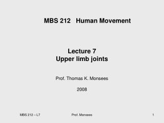
MBS 212 Human Movement. Lecture 3 Upper limb movements 1/2 Prof. Thomas K. Monsees 2008. Upper limb. L3 + 4: Upper limb movements L5 + 6: Forearm and hand movements L7: Upper limb joints. Pectoral Girdle. Pectoral girdle attached upper limb to trunk

MBS 212 Human Movement
E N D
Presentation Transcript
MBS 212 Human Movement Lecture 3 Upper limb movements 1/2 Prof. Thomas K. Monsees 2008 Prof. Monsees
Upper limb • L3 + 4: Upper limb movements • L5 + 6: Forearm and hand movements • L7: Upper limb joints Prof. Monsees
Pectoral Girdle • Pectoral girdle attached upper limb to trunk • Only point of articulation (!) with axial skeleton is the sterno clavicular joint • Scapula rides in a sea of muscles attaching it to head, neck + thorax (rotator cuff) • Clavicle acts as a strut holding upper limb away from trunk Sterno clavicular joint Prof. Monsees
Upper limb: Overview Bones Anterior view • Pectoral girdle • Scapula, Clavicle • Arm • Humerus • Forearm • Radius, Ulna • Hand • Carpus, Metacarpus, Phalanges Superior view Prof. Monsees
Scapula • Large, flat, triangular bone • Situated in posterolateral aspect of thorax • Overlapping 2nd-7th ribs • Suspended in muscles (rotator cuff) Prof. Monsees
Bony landmarks • Acromion process • Forms protective cover • Attachment for clavicle • Attachment for muscles • Coracoid process • Attachment for muscles • Glenoid cavity • Articulates with humerus • Infraglenoid tubercle • Att. long h. triceps brachii • Spine • Att. trapezius, deltoid posterior posterior • Angels: superior, lateral, inferior • Borders: medial, lateral, superior Prof. Monsees
lateral Bony landmarks • supraglenoid tubercle • Att. long h. biceps brachii • Subscapular fossa • Att. subscapularis Prof. Monsees
Scapula articulates with: • Clavicle at acromion process forming acromioclavicular joint • Head of humerus at glenoid fossa forming shoulder joint (gleno humeral joint) Prof. Monsees
Clavicle • Curved bone • Smooth superior surface, roughened inferior • Expanded medial, flattened lateral ends • Articulates laterally with scapula (acromio clavicular joint) • medially with sternum forming sterno clavicular joint Prof. Monsees
Humerus Proximal end • Long bone of arm • Head articulates with gleonid cavity of scapula • greater tubercle, att. of • Supraspinatus (superior) • Infraspinatus (middle) • Teres minor (inferior) • lesser tubercle, att. of • subscapularis • Intertubercular sulcus • tendon long head biceps brachii • Roughened shaft surface for att • Pectoralis major, teres major, latissimus dorsi, deltoid, coracobrachialis Prof. Monsees
Humerus • flattened distal ends with epicondyles (med. + lat.) • Capitulum articulates with head of radius • Trochlear articulates with trochlear notch of ulna (elbow joint) distal end Prof. Monsees
Articulations or Joints • Articulation or Joint • Place where two bones come together • Structure correlated with movement • Named • According to bones or parts united at joint • According to only one of articulating bones • By Latin equivalent of common name Prof. Monsees
Joints: classification by function • i.e., by the degree of movement possible: • Synarthroses • Joints with little or no movement • Skull sutures, mental symphysis, teeth in sockets, 1st costosternal joint. • Amphiarthroses • Slightly moveable joints • Intervertebral discs, costosternal joints, pubic symphysis • Diarthroses • Freely moveable joints • Shoulder, knee, hip, elbow, interphalangeal, tarsal, and carpal joints Prof. Monsees
Joints: classification by structure Prof. Monsees
Structural joint classification • Bony fusion: (synostosis), fusion of 2 bones, boundary between disappears, totally rigid, e.g. metopic suture of paired frontal bones of the skull • Cartilaginous joints: Bones held together by cartilage; no joint cavity Epiphyseal plates of long bones, costosternal joints, pubic symphysis, intervertebral discs Metopic suture at birth Prof. Monsees
Joint Classification 3. Fibrous joints: • Bones held together by collagenous fibers extending from the matrix of one bone into the matrix of the next. • No joint cavity • Skull sutures, teeth in joints, distal radioulnar joints & tibiofibular joints Prof. Monsees
Fibrous joints (cont..) Syndesmoses Gomphoses Gomphosis Prof. Monsees
Joints: classification by structure 4. Synovial joints: • Bones separated by a joint cavity; lubricated by synovial fluid; enclosed in a fibrous joint capsule. • Name some synovial joints! • Shoulder, hip, elbow, knee, carpal, interphalangeal Prof. Monsees
Sagittal suture Lamboid suture Structure and Function • Joints are designed for their function. • Let’s look at sutures as our 1st example: • Connective tissue? • Degree of Movement? • What kind of joint? • Function? • Fibrous joint • Synarthroses • skull bone moveable at birth • but rigid later Prof. Monsees
Structure and Function • Let’s look at some symphyses. • What kind of joint is a symphysis? What kind of movement is possible? • Name a symphysis! (an obvious one is in the picture) • What connects the bones in these joints? Prof. Monsees
Structure and Function • Now let’s talk about synovial joints. • How do they differ from the previous 2? • 5 main structural characteristics: • Articular cartilage • What kind of cartilage is it? (H _ _ _ _ _ _ ) • Where do we find it? • What does it do? Prof. Monsees
Structure and Function • Articular capsule • 2 layered. surrounds both articular cartilages and the space btwn them. • External layer is made of dense irregular CT & is continuous with the perisoteum. • Inner layer is a synovial membrane made of loose connective tissue. • It covers all internal joint surfaces except for those areas covered by the articular cartilage. Prof. Monsees
Structure and Function • Joint(Synovial) Cavity • The potential space within the joint capsule and articular cartilage • Synovial Fluid • A small amount of slippery fluid occupying all free space w/i the joint capsule • Formed by filtration of blood flowing thru capillaries in the synovial membrane • Synovial fluid becomes less viscous as joint activity increases. • Lubrication • Nutrient distribution • Shock absorption Prof. Monsees
Structure and Function • ReinforcingLigaments • Support, strengthen, reinforce synovial joint • These ligaments are very strong: with excessive force, one of the attached bones usually breaks before the ligament tears • Collagen fibers Prof. Monsees
Other Synovial Structures • The knee and hip joints have cushioning fatty pads btwn the fibrous capsule and the synovial membrane or bone. • Discs of fibrocartilage (i.e., menisci) which improve the fit btwn bone ends, thus stabilizing the joint. • Found in the knee, jaw, and sternoclavicular joint. • Bursae are basically bags of lubricant - fibrous membrane bags filled w/ synovial fluid. Often found where bones, muscles, tendons, or ligaments rub together. Prof. Monsees
Structural classification of synovial joints • Synovial joints are described as gliding, pivot, saddle etc on the basis of the shape of the articulating surfaces • Each type of joint permits a different type and range of motion Prof. Monsees
Types of Synovial Joints • Planejoints (gliding) • Articular surfaces are flat and allow short slipping or gliding movements. • Slight nonaxial or multiaxial • Intercarpal and intertarsal joints • Hinge joints • A cylindrical projection of one bone fits into a trough-shaped surface on another (like a hotdog in a bun) • Movement resembles a door hinge. • monaxial • Elbow joint – ulna and humerus; Interphalangeal joints Prof. Monsees
Type of Synovial Joints • Pivot joints • Rounded end of one bone protrudes into a ring formed by another bone or by ligaments of that bone. • Monaxial (rotation) • Proximal radioulnar joint • Atlas-axial joint • Condyloid joints (ellipsoidal) • Oval articular surface of one bone fits into a complementary depression on another. • biaxial • Radiocarpal joints • Metacarpophalangeal joints Prof. Monsees
Types of Synovial Joints • Saddle joints • Each articular surface has convex and concave areas. Each articular surface is saddle-shaped. • biaxial • Carpometacarpal joints of the thumbs. • Ball-and-Socket joints • Spherical or semi-spherical head of one bone articulates with the cuplike socket of another. • Allow for much freedom of motion. • triaxial • Shoulder and hip joints. Prof. Monsees
Movements: Definitions Movements are described with respect to the anatomical position as reference Sagittal • What is the anatomical position? • Standing upright • Feet together • Hands by the side • Face looking forward • Anatomical planes? • Coronal (vertically) anterior + posterior parts of body • Sagittal (vertically, median) right + left parts • Transverse (horizontal, medial) superior + inferior parts Coronal Transverse Prof. Monsees
Flexion - Extension Arm movement • Flexion • Movement anterior to coronal plane, angle between bone decreases • Extension • Moving posterior to coronal plane, angle between bone increases (straightening) • Also movement beyond anatomical position in direction opposite to flexion Bending of trunk Flexing neck to chest Prof. Monsees
Abduction and Adduction • Abduction (abroad and away…) • movement which brings a limb - arm or leg – more away from the sagittal plane of the body • Adduction • movement which brings a limb closer to the sagittal plane of the body Prof. Monsees
Protraction and Retraction Retraction: posterior movement Protraction: anterior movement Prof. Monsees
Movements of scapula Abduction(up) Adduction(down) • Upper limb is highly mobile for positioning hand in space • Scapula can move relative to trunk • Protraction + retraction (sliding) • Abduction + adduction (rotation) • Shoulder (glenohumeral) joint allows arm to move around 3 axis • Flexion + extension • Abduction + adduction • Medial + lateral rotation • circumduction • Retraction(back) • Protraction(front) Prof. Monsees
Movements of arm at shoulder joint • Abduction • Adduction • Flexion • Extension • Medial rotation • Lateral rotation • Circulation Prof. Monsees
Muscles of the shoulder girdle • Bind shoulder girdle to axial skeleton • M. trapezius, m. levator scapulae, m. serratus anterior, m. pectoralis minor • Attach humerus to axial skeleton • M. pectoralis major, m. latissimus dorsi • Bind humerus to shoulder girdle • M. teres major, m. deltoideus, rotator cuff muscles • Muscles of anterior/ posterior compartment of upper arm • M. biceps brachii et brachialis/ m. triceps brachii Prof. Monsees
Overview: Muscles of the shoulder Prof. Monsees
Overview: Rotator cuff muscles Prof. Monsees
Muscles of the Arm L 5 + 6 Prof. Monsees
Nerve supply muscles upper limbbrachial plexus (bp) • All major nerves innervating upper limb originates from brachial plexus • Bp formed by anterior rami of C5 to C8 and T1 • Pt: roots, trunks, divisions, and cords Prof. Monsees
Nerve supply muscles upper limbbrachial plexus Axillary artery Median nerve Prof. Monsees Radial nerve Ulnar nerve
Muscles that bind shoulder girdle to axial skeleton • M. trapezius • M. levator scapulae • M. serratus anterior • M. pectoralis minor Thank you for your attention! Prof. Monsees