Download
1 / 30
300 likes | 568 Vues
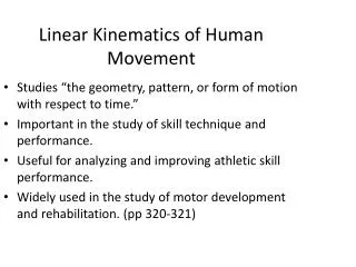
MBS 212 Human Movement. PRACTICAL 3 LOWER LIMB Prof. Thomas K. Monsees 2008. Regions of the lower limb. Gluteal Posterolateral btw iliac crest + gluteal fold of buttock Thigh Anteriorly btw inguinal lig + knee joint Leg Btw knee + ankle joint Foot Distal to ankle point.

E N D
MBS 212 Human Movement PRACTICAL 3 LOWER LIMB Prof. Thomas K. Monsees 2008 Prof. Monsees
Regions of the lower limb • Gluteal • Posterolateral btw iliac crest + gluteal fold of buttock • Thigh • Anteriorly btw inguinal lig + knee joint • Leg • Btw knee + ankle joint • Foot • Distal to ankle point Prof. Monsees
Bones of the lower limb Bones • Pelvic bone (link to sacrum) • Femur (largest bone of body) • Tibia (medial) • Fibula (lateral) Joints • Hip joint (ball & socket) • Btw pelvic bone + femur • Knee joint w patella (hinge) • Btw femur + tibia Prof. Monsees
Transition from abdomen and pelvis to lower limb Bony pelvis, lat view Prof. Monsees
Proximal end of right femur Prof. Monsees
distal pt Femur Prof. Monsees
Tibia and fibula Prof. Monsees
Bones of the foot Bones • Phalanges • Metatarsals • Tarsal bones • 7 in 2 rows + 1 bone in between Joints Ligaments Deep transverse metatarsal ligament Prof. Monsees
Bones of foot do not lie flat in single plane • Longitudinal + transverse arches of the foot • Arches are flexible; absorb and transmit forces during standing and walking Prof. Monsees
Hip Muscles • Muscles in gluteal region mainly support extension, rotation + abduction of hip • They also control movement of pelvis relative to limb bearing the body’s weight (stance limb) while the other limb swings forward (swing limb) during walking Prof. Monsees
Hip Muscles • Major flexors of hip attach to posterior abdominal wall and descend through gap between inguinal ligament and pelvic bone to attach to proximal end of femur Prof. Monsees
Thigh Muscles • Separated into 3 compartments by fascia, bones + ligaments • Medial (adductor) • Anterior (extensor) • Posterior (flexor) • Most medial muscles act mainly on hip joint • Large posterior hamstring muscles act on hip (extension) + knee (flexion) • Anterior muscles (quadricaps femoris) extend knee Prof. Monsees
Leg Muscles • Separated into 3 compartments by fascia, bones + ligaments • Lateral (fibular) • Anterior • Posterior • Lateral muscles predominantly evert foot • Anterior muscles dorsiflex foot + extent digits • Posterior muscles plantarflex foot + flex digits Prof. Monsees
Right posterior hip, superficialdeep Prof. Monsees
Right anterior thigh Right medial thigh Prof. Monsees
Right posterior thigh Prof. Monsees
Cross section left leg Prof. Monsees
Anterior view right legLateral view right leg Prof. Monsees
Posterior view right leg Posterior view right leg Calf muscle, superficial Calf muscle, deep Prof. Monsees
Right foot musclessuperficial deep Prof. Monsees
Surface anatomy lower limb Prof. Monsees
Femor triangle Wedge-shaped depression formed by muscles in the upper thigh btw anterior abdominal wall + lower limb Content: Major blood supply Femoral nerve Prof. Monsees Boundaries
Popliteal fossa: • Posterior to knee joint • Diamond shaped region formed by muscles of thigh + leg • major vessels + nerves pass here • Tarsal tunnel: • series of cannels on posterior side of ankle. Most nerves, vessels + flexor tendons pass here between leg and foot • Cannel formed by adjacent bones + flexor retinaculum Prof. Monsees
Adductor canal Prof. Monsees