Download
1 / 62
620 likes | 752 Vues
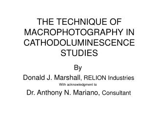
THE TECHNIQUE OF MACROPHOTOGRAPHY IN CATHODOLUMINESCENCE STUDIES. By Donald J. Marshall , RELION Industries With acknowledgment to Dr. Anthony N. Mariano, Consultant. MACROPHOTOGRAPHY. MICRO vs MACRO CLASSICAL DEFINITION IS A PHOTO BIGGER THAN LIFE

E N D
THE TECHNIQUE OF MACROPHOTOGRAPHY IN CATHODOLUMINESCENCE STUDIES By Donald J. Marshall, RELION Industries With acknowledgment to Dr. Anthony N. Mariano, Consultant
MACROPHOTOGRAPHY • MICRO vs MACRO • CLASSICAL DEFINITION IS A PHOTO BIGGER THAN LIFE • DOESN’T WORK FOR MICROSCOPISTS BECAUSE ALL PHOTOS ARE LIKE THIS • SO MACROPHOTOGRAPHY MEANS, TO US, ONLY CAMERA IS USED AND LARGE SPECIMEN AREA IS VIEWED
MACROPHOTOGRAPHY • Used only by a few investigators • Microscope with very low magnification objective (1X) sometimes used; • But usually no microscope– only a camera. • Viewed area 2 to 5 cm in diameter
WILLIAM CROOKES, F.R.S. • 1880. Philosophical Transactions of Royal Society • “….preparation of sulphide of calcium……. shines with a bright blue-violet light, and, when on a surface of several square inches, is sufficient to light up a room……. ”
MACRO vs MICRO • Unfocused cold cathode beam about 1 cm. diameter. • Top window in some cases limits viewed area to 7 to 8 mm. • So arbitrary dividing line is: • 5 mm or less is micro. • 5 mm to 5 cm or greater is macro.
OPERATIONAL REQUIREMENTS • Capability to defocus the electron beam. • Sufficient electron beam current to provide viewable cathodoluminescence. • Large viewing window area.
FOCUS and DEFOCUS • Unfocused beam implies that nothing is done to the beam to change its size. • Unfocused beam is usually not large enough in diameter – only 0.5 cm to 1 cm. • So defocusing is done.
RASTERING • Could also be done, in principle, by rastering beam but instrumentation requirements are more involved and this is not done on simple cold cathode-based instruments
FOCUSING AND DEFOCUSING OF ELECTRON BEAM • Requires special lens element in electron gun to defocus electron beam • Both electrostatic and electromagnetic lens are possible. • Electromagnetic lens is most common
FOCUSING AND DEFOCUSING • In normal operation, • focus point moves closer to coil (stronger focus) with higher coil current and • Focus point moves away from coil (weaker focus) with lower coil current.
DEFLECTION, FOCUSING, AND ABERRATIONS • Coils are usually designed for the best small spot size and shape. • Coils are not designed for best defocused spot shapes and there is some appreciable variation in results. • The spot can always be made larger but the shape may be less than circular or elliptical and some compromise is necessary. • For those instruments which include magnetic deflection of the beam, there is an inevitable interaction between the deflection and focusing systems. • Any deviations from the ideal beam shapes are termed aberrations. If there were sufficient demand for macro CL work, then coil designers would rise to the occasion.
DEFOCUSED BEAM SHAPE EXAMPLE • Scottish Sandstone • Beam is large but it is strongly elliptical 5 cm
Dr. James Clark • Macro CL used originally to examine panned concentrates from stream sediments in carbonatite exploration. • Used in granitoids where examination of larger areas facilitates identification of feldspar phase relationships and pathways for meteoric alteration fluids. • Also to determine timing of quartz and carbonate events associated with gold mineralization and examination of cross cutting relationships in veins. • Dr. James Clark, Applied Petrographics. • http://www.appliedpetrographics.com
GRANODIORITICINTRUSIVE ROCK • K feldspar - light blue CL - weak reddish purple overprint. Unaltered plagioclase - yellowish green overprinted by reddish brown to light brown CL with progressively stronger clay alteration. • The K feldspar poikilitically encompasses smaller crystals of plagioclase. • Quartz and micas are non-luminescent. Field of view = 15.42 mm. • Courtesy of Dr. James Clark
GRANODIORITIC INTRUSIVE ROCK • K feldspar light blue CL. Unaltered plagioclase yellowish green CL • The K feldspar poikilitically encompasses smaller crystals of plagioclase. • Quartz and micas are non-luminescent. • Field of view= 15.42 mm. • Courtesy of Dr. James Clark 1. 5 centimeter
MULTIPLE EPITHERMAL QUARTZ TYPES • Epithermal quartz vein with CL • CL photo shows two varieties of microcrystalline quartz (tan and dull red), while the comb-textured quartz has fine-scale growth zoning in shades of yellow, red, gray, and blue CL. • Dr. James Clark 1.48 cm
COMPLEX BANDED Q-CHALCEDONY VEIN LATE STAGE CHALCEDONY AND QUARTZ • Complex epithermal Q-chalcedony vein. CL photo highlights a quartz vein stratigraphy with early non-luminescent microcrystalline quartz (Q1), an intermediate stage of microcrystalline quartz with moderate yellowish-brown CL, and late-stage chalcedony and quartz with strong yellow CL (Q3). The main vein is cut by a narrow veinlet of Q2 and orange-luminescent calcite. • Dr. James Clark EARLY NON-LUMINESCENT QUARTZ CALCITE 2.14 cm
DRUSY QUARTZ VEIN • Drusy quartz in a polished slab of quartz vein material from an epithermal silver-gold mine, Durango, Mexico. Locally this quartz has native gold inclusions. No zonation visible in plane light. • Kodak Royal Gold 200 film, 60 sec. 12.5 kV, 0.6ma. • Vertical dimension is 19 mm. • Dr. James Clark 1.4 cm
Quartz-calcite-adularia vein • Quartz-adularia-calcite vein. Lattice- and parallel-bladed acicular calcite needles with interstitial quartz-adularia. Bright orange calcite overwhelms much weaker brown CL of adularia-quartz assemblage 1.54 cm
Quartz-calcite-adularia vein • Quartz-calcite-adularia vein. A band of lattice-bladed calcite has interstitial microcrystalline quartz-adularia. Microdrusy quartz partly lines voids within intersecting calcite blades. Calcite has bright orange CL and overwhelms the much subtler CL of adularia. The microdrusy quartz has zoned yellow CL (appears green in the photo). • CL; field of view= 11.57mm. • Courtesy of Dr. James Clark. 2 mm 1.2 centimeter
DEWEY LIMESTONE • Devonian from Oklahoma • Macro provides a quick picture of the different major phases and serves as a guide to more detailed examination 5 cm.
CORAL • Solitary coral, Siphonophyllia Sp., Co. Sligo, perpendicular to long dimension. • 14 kV, 1 m A, 2 seconds • Sample from Claire Mulhall, Trinity College, Dublin 5 cm.
2 mm 5 cm.
CORAL • Solitary coral, Siphonophyllia Sp., Co. Sligo, parallel to long dimension. • 14 kV, 1 mA, 2 seconds • Sample from Claire Mulhall, Trinity College, Dublin 5 cm.
LIMESTONE DRILL CORE • Partially dolomitised Waulsortian Limestone, Co. Tipperary • 14 kV, 1 mA, 2 sec. • Sample from Claire Mulhall, Trinity College, Dublin 5 cm.
LIMESTONE DRILL CORE • Limestone drill core expanded X2 to emphasize dolomite crystals 2.5 cm.
MAP FOR MICRODRILLING • Jay Kaufman, U. Md. • Massive to finely laminated carbonate from Namibia. • Determined Mn/Sr, 87Sr/86Sr,C and O isotopes from small selected areas. • Kaufman et al., 1991, Precambrian Research, 49, p. 301 • Kaufman et al., 1993, Earth and Planetary Science Letters, 120, p.409
MICRODRILLING OF LAMINATED CARBONATE • MLM = moderately luminescent microspar • LSLC = luminescent sparry calcite • NLM = non-luminescent microspar • WR = whole rock
Map for microdrilling • Jay Kaufman et al from proterozoic in Namibia. • Drilled out samples from 1 mm diameter areas for C and O isotopic analyses • WR = whole rock value. Local values shown also 2.9 cm
Map for microdrilling • Jay Kaufman et al, • Drilled out 1 mm areas for Mn/Sr and 87Sr/86Sr analyses. • WR = whole rock value. Local values shown also 2.9 cm
Dr. Anthony N. Mariano • Specialist in mineral deposits of all kinds and especially those associated with rare earths. Investigated them all over the world on five continents. • Has been using CL for more than 30 years. • Dr. Anthony N. Mariano, Carlisle, MA USA
CARBONATITES • Dr. Mariano uses macrophotos extensively in his work on carbonatites and especially likes to work with slabs. • “Slabs give you a better picture of what the rock really looks like and especially as an initial overall look.” (Dr. Anthony Mariano)
Apatite Søvite – Okorusu, Namibia • Light lilac – pink CL apatite – LREE activated • Orange CL calcite, Mn2+ • Non-CL - pyroxene • 17 kV,0.8 mA • All apatite in carbonatite are LREE 46 millimeters
Søvite, Okorusu, Namibia • Various stages of calcite crystallization showing variations in orange to orange-red CL, corresponding to changes in trace element content of calcite with each crystallization episode. • Also blue fluorite 46 mm
Nepheline Syenite, Okorusu, Namibia • Light blue CL plagioclase • Grey (non-CL) nepheline • Dull red CL specks are sodalite • Not a carbonatite 46 mm in length
Fluorite ore and apatite, Okorusu, Namibia • Hydrothermal mineralization of fluorite and apatite • light blue – zoned fluorite, intrinsic • light lilac-pink apatite, LREE • Really dark is quartz or voids. • Dr. Anthony N. Mariano 46 mm
Carbonatite, Matongo Bandaga, Burundi • Blue apatite, LREE activators • Bright red, fenite Fspars (Fe3+) • dark areas are blue – non-luminescing pyroxenes (aegerine) • Orange calcite • Dr. Anthony N. Mariano 46 mm
Contact between apatite – magnetite søvite and fenite, Okorusu, Namibia • Fenite carbonatite contact • knob on upper right side is Søvite with orange CL calcite, light blue CL apatite and non-CL magnetite. Band on left is dominant orange CL calcite that drowns out red, Fe3+ activated CL, of K feldspar. In plane light band is leucocratic and in sharp contact with central melanocratic band. The light colored matrix is calcite and orthoclase and the dark green disseminated grains are diopside. • The central band contains non-CL diopside and veins of dull grey CL celsian (activator not known). First reported occurrence of celsian (BaAlSi3O8) in carbonatite. • Late bands are calcite • 16 kV, 0.9 mA, 30 seconds, ASA • Dr. Anthony N. Mariano 46 mm
GEMSTONES • Not surprisingly, gem stones are one of the favorite candidates for CL macrophotograpy. • They are often large. • They usually have an irregular, non-planar, shape. • Often they are not removable from their mounts
SYNTHETIC DIAMOND • Synthetic diamond CL at room temperature • Dr. C.M. Welbourn, Diamond Sales, Ltd. 5.1 millimeters
SYNTHETIC DIAMOND • Synthetic diamond CL at low temperatures, 80 degrees K • Dr. C. M. Welbourn, Diamond Sales, Ltd. 5.1 millimeters
Dr. Johann Ponahlo • Physical-chemist and gemologist. • Has used CL macro macro techniques for description and documentation of natural and synthetic gemstones for more than two decades. • Dr. Johann Ponahlo, Vienna, Austria
“Samples of considerable thickness and/or of irregular shape can be studied as well as specimens in their original settings and even small figurines. “ Dr. Johann Ponahlo