Download
1 / 15
220 likes | 1.18k Vues
Cathodoluminescence of quantum dots. IMAIZUMI Kei Itoh Lab. CONTENTS. 1. Quantum dot. ・ Quantum dot ・ Cathodoluminescence. 2. CdSe quantum dots. ・ CdSe quantum dots. Ref ) I. Yamakawa et al . J. Luminescence 87-89 (2000) 384-386. 3. My study - CuCl nanoparticles.

E N D
Cathodoluminescenceof quantum dots IMAIZUMI Kei Itoh Lab.
CONTENTS 1. Quantum dot ・Quantum dot ・Cathodoluminescence 2. CdSe quantum dots ・CdSe quantum dots Ref) I. Yamakawa et al. J. Luminescence 87-89 (2000) 384-386. 3. My study - CuCl nanoparticles ・CuCl quantum dots
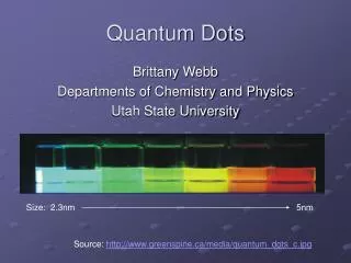
Quantum dots (QDs) Quantum dots Energy level electron confinement Quantum size effect ・Discrete energy levels (artificial atom) ・Energy shift depends on the size The character of QDs depends on the dot size
Size dependence (Example) Ref) http://quantumdot.lanl.gov/ The color depends on the size ・investigating individual dot ・size selection The way to look at the single particle?
light 100nm~ ~10nm Diffraction limit and microscope The beam can be focused only about the half of the wavelength. Diffraction limit electron microscope optical microscope electron beam
Electron beam Cathodo- luminescence Secondary electron →SEM Continuous X-ray Characteristic X-ray →EDS Sample Cathodoluminescence (CL) It is possible to measure CL spectrum with SEM image. SEM system (Itoh Lab.) SEM : Scanning Electron Microscope EDS : Energy Dispersive X-ray Spectroscopy
CdSe dots embedded in ZnSe Ref) I. Yamakawa et al. J. Luminescence 87-89 (2000) 384-386. Many groups reported about CdSe dots growth, characterization etc… Various conclusions structure, morphology size, composition etc… The authors investigated the character of CdSe dots.
Sample & TEM Image Dot Sample CdSe dots embedded in ZnSe CdSe ZnSe 10nm ZnSe 50nm GaAs Ref) S.V.Ivanov et al. Appl. Phys. Lett. 74 (1999) 498. Dot size : 15~40nm Dot density : 2×10¹⁰ /cm2 TEM : Transmission Electron Microscope
CL and PL measurement sharp spikes [CL] beam spot (diameter 200nm) T = 4.2K CL spectrum PL spectrum CdSe nanostructure? PL : Photoluminescence
Estimating the dot density Simulation CL Spectrum dot density ~5×10¹¹/cm2 smaller size dots (invisible by TEM) (2×10¹⁰ /cm2 (TEM)) 2type size dots Further calculation revealed the existence of the 6~10nm CdSe dots. 6~10nm 15~40nm
1 cm My Study – CuCl nanoparticles CuCl Tablet It was made by pressing the CuCl powder. CuCl nanoparticles Pulse laser Laser ablation CuCl nanoparticles
PL measurement PL spectrum reflects the luminescence from all particle T = 2K SEM Image (10nm~10μm particle)
CL measurement T = 15K SEM Image The spectrum of ~100nm CuCl particle has been observed.
Under researching ? 15nm We are measuring the spectrum of smaller dots. Discussing the difference with bulk (quantum size effect)
SUMMARY Because the character of quantum dot depends on the size, it is important to investigate the single dot. Cathodoluminescence measurement is the powerful tool to look at the small dots. We are measuring the CL spectrum of CuCl nanoparticle (about 10nm).