Duchenne Muscular Dystrophy
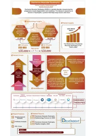
Duchenne Muscular Dystrophy. PG SBI3U. Introduction. Duchenne Muscular Dystrophy (DMD) is the most common and most serious form of muscular dystrophy 1 in 3500 boys are affected DMD is a fatal genetic disorder that damages muscle tissue

Duchenne Muscular Dystrophy
E N D
Presentation Transcript
Duchenne Muscular Dystrophy PG SBI3U
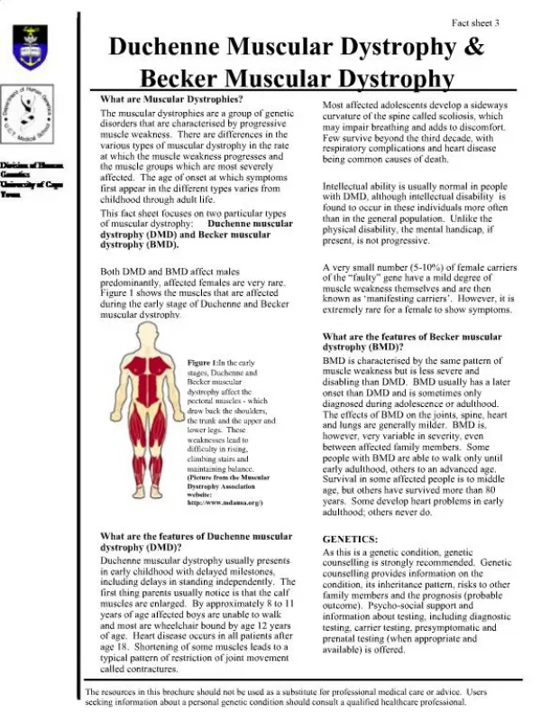
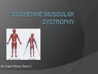
Introduction • Duchenne Muscular Dystrophy (DMD) is the most common and most serious form of muscular dystrophy • 1 in 3500 boys are affected • DMD is a fatal genetic disorder that damages muscle tissue • DMD first affects large skeletal/voluntary muscles, weakening the arms and legs, but eventually progresses to affecting all muscle groups
History • Sir Charles Bell first described muscular dystrophy in 1930 • DMD named for Guillame Benjamin Amand Duchenne • Wrote about the disease from 1861 to his death in 1875 • Called it “paralysie musculaire pseudohypertrophique” which means muscular paralysis with swollen muscles due to fat replacing muscle tissue • The gene for DMD was located on the X-chromosome between 1978 and 1983 • In 1985 DNA markers were identified for carrier and prenatal diagnosis • Between 1987 and 1988 the gene responsible for DMD was cloned and its protein product (dystrophin) was identified
Cause • A genetic defect in X chromosome • Mutation in gene for the protein dystrophin • The largest known human gene • Protein in the cell membrane of muscles • Its absence causes DMD, if the protein is partially functional Becker’s MD results • Causes creatine kinase to leak from the cell and excess calcium to enter into muscle cells • The cells die and are replaced by fat or connective tissue
Inheritance • Almost exclusive to boys, very few girls have it • The mutant gene for DMD is recessive and located on the X-chromosome • Girls only get DMD if their father has DMD and their mother is a carrier • 1/3 are a new mutation, not inherited
Effects • First manifests as a toddler • Half start walking later than normal (18 months) • Babies have muscle degradation but grow/regenerate fast enough to make up for the damage • Toddlers have difficulty walking, getting up or climbing stairs • May not be able to run or jump
Effects (cont.) • Enlarged calves, muscle is replaced with fat/connective tissue • Called pseudohypertrophy • Stand up with the “Gower maneuver” • Fatigue • 1/3 have mild to moderate mental impairment
Effects (cont.) • Ages 6-9 walk on toes, with belly out, chest back, legs wide apart • Muscle contractures of Achilles tendon and hamstrings • Most affected boys lose the ability to walk by age 12 • Inactivity may cause scoliosis, osteoporosis or muscle atrophy
Effects (cont.) • Late Stages of Disease: • Heart and diaphragm are weak • Difficulty breathing, a tracheotomy tube or ventilator may be necessary • Most develop a dilated cardiomyopathy • Most affected people die before age 30 due to heart failure, lung failure or respiratory infections Tracheotomy tube
Diagnosis • Distinctive swelling in calves, use of “Gower maneuver” • High levels of creatine kinase in blood • Electromyography • Genetic testing • Muscle biopsy • MRI or electron microscopy can determine extent of muscle damage • Can be diagnosed prenatally (amniocentesis) • Carriers can also be diagnosed: • 60-70% have high levels of creatine kinase • 5% have some form of muscle weakness • Genetic analysis
Treatment • No cure • Physical therapy, corticosteroids, immunosuppressive and antibiotic drugs, and mechanical aids prolong mobility • Swimming is a common physical therapy • Inactivity (bedrest) worsens the disease • A high-fibre, high-protein, low calorie diet is recommended
Future • Potential treatments: • Gene therapy • To replace faulty dystrophin gene • To add an extra utrophin gene • To “turn off” myostatin gene • Exon skipping • Stem cell or myoblast transplants • Drugs already used for other disorders • HCT1026 (arthritis) • Losartan (high blood pressure)
References • Abramovitz, M. (2008). Muscular Dystrophy. Farmington Hills, Michigan: Gale Cengage Learning. • Bunch, B. (2003). Muscular Dystrophy. In Diseases (Volume 6, pp. 5-8). Dunbury, CT: Scientific Publishing, Inc. • Colapinto, J. (12/20/2010). Mother Courage. New Yorker, Vol. 86 Issue 41. Retrieved May 8, 2011 from http://web.ebscohost.com/ehost/detail?vid=7&hid=14&sid=6d5e49c8-1096-4d56-ac0f-82ab0227b694%40sessionmgr4&bdata=JnNpdGU9ZWhvc3QtbGl2ZQ%3d%3d#db=lfh&AN=55984630 • Emery, A.E.H. (2008). The Facts: Muscular Dystrophy. Oxford: Oxford University Press • Medline Plus. (2010). Duchenne Muscular Dystrophy. Retrieved May 8, 2011 from http://www.nlm.nih.gov/medlineplus/ency/article/000705.htm • National Institute of Neurological Disorders and Stroke. (2011). Muscular Dystrophy: Hope Through Research. Retrieved May 11, 2011 from http://www.ninds.nih.gov/disorders/md/detail_md.htm • Parent Project Muscular Dystrophy. (2010). Retrieved May 9, 2011 from http://www.parentprojectmd.org • PubMed Health. (2010). Duchenne Muscular Dystrophy. Retrieved May 8, 2011 from http://www.ncbi.nlm.nih.gov/pubmedhealth/PMH0001724/ • Wynbrant, J. (2000) Muscular Dystrophy.In The Encyclopedia of Genetic Disorders and Birth Defects (pp. 231). New York: Facts on Fire, Inc.
References (Images) • Slide 5: http://drugster.info/img/ail/459_462_3.jpg • Slide 7 (top): http://neuromuscular.wustl.edu/pics/people/patients/beckerl.jpg • Slide 7 (bottom): http://www.parentprojectmd.org/images/content/pagebuilder/GowerManeuverLarge.jpg • Slide 8 (top): http://drugster.info/img/ail/459_462_2.gif • Slide 8 (bottom): http://kwikfit4u.com/kwikblog/wp-content/uploads/2010/09/scoliosis.jpg • Slide 9 (top): http://www.vnacarenewengland.org/healthGate/images/tracheostomy_tube.jpg • Slide 9 (bottom): http://withfriendship.com/images/h/35958/dilated-cardiomyopathy.jpg • Slide 11: http://www.homecarespecialistsinc.com/images/medequipment/Wheelchair.jpg • Slide 12 (top): http://library.thinkquest.org/28000/media/genetherapy/l_gene.therapy-ms.gif • Slide 12 (bottom): http://www.nature.com/nrg/journal/v4/n10/images/nrg1180-f2.gif