Download
1 / 109
1.14k likes | 1.52k Vues
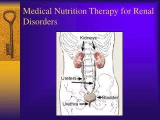
Medical Nutrition Therapy for Renal Disorders. Functions of the Kidney. Excretory Acid-base balance Endocrine Fluid and electrolyte balance. Excretory Functions. Removal of excess fluid and waste products 180 L of filtrate pass through the kidneys each day producing 1-2 L of urine

E N D
Functions of the Kidney • Excretory • Acid-base balance • Endocrine • Fluid and electrolyte balance
Excretory Functions • Removal of excess fluid and waste products • 180 L of filtrate pass through the kidneys each day producing 1-2 L of urine • Wastes excreted from the body in urine include urea (byproduct of protein metabolism); excess vitamins and minerals; metabolites of some drugs and poisons
Acid-Base Functions • Acid-base balance is maintained through a buffer system, which maintains blood at pH of 7.4 • Bicarbonate carries hydrogen ions to the kidneys where they are removed from extracellular fluid in the tubules, returned to the bloodstream as needed • Phosphate buffers intracellular fluid Source: Byham-Gray, Wiesen, eds. A Clinical Guide to Nutrition Care in Kidney Disease. ADA, 2004
Acid-Base Balance Functions • When fluid volume is low, anti-diuretic hormone (ADH) or vasopressin is released from the anterior pituitary; increases absorption of water in the collecting duct • When extracellular volume (ECV) decreases, the renin-angiotensin-aldosterone system is activated excretes less sodium chloride Source: Byham-Gray, Wiesen, eds. A Clinical Guide to Nutrition Care in Kidney Disease. ADA, 2004
Endocrine Functions • 1,25-dihydroxy-vitamin D3 or calcitriol is produced in the kidney; enhances calcium absorption • Activation of Vitamin D and excretion of excess phosphate maintain healthy bones • Erythropoietin: acts on the bone marrow to increase production of red blood cells Source: Byham-Gray, Wiesen, eds. A Clinical Guide to Nutrition Care in Kidney Disease. ADA, 2004 Source: Byham-Gray, Wiesen, eds. A Clinical Guide to Nutrition Care in Kidney Disease. ADA, 2004
The Most Common Kidney Diseases • Diabetic Nephropathy damage to the nephrons in the kidneys from unused sugar in the blood, usually due to Diabetes. • High Blood Pressure can damage the small blood vessels in the kidneys. The damaged vessels cannot filter poison from the blood as they are supposed to. • Polycystic Kidney Disease (PKD) is a hereditary kidney disease in which many cysts grow in the kidneys. These cysts may lead to kidney failure.
The Most Common Kidney Diseases • Acute Renal Failure - Sudden kidney failure caused by blood loss, drugs or poisons. If the kidneys are not seriously damaged, acute renal failure may be reversed. • Chronic Renal Failure - Gradual loss of kidney function is called Chronic Renal Failure or Chronic Renal Disease. • End-Stage Renal Disease - The condition of total or nearly total and permanent kidney failure.
Kidney Diseases • Glomerular diseases • Nephrotic syndrome • Nephritic syndrome—tubular or interstitial • Tubular defects • Acute renal failure (ARF) • Other • End-stage renal disease (ESRD) • Kidney stones
Nephrotic Syndrome • Alterations of the glomerular basement membrane allows persistent loss of large amounts of protein in the urine • Associated with diabetes, glomerulonephritis, amyloidosis, lupus • High risk for cardiovascular disease • Hypercoagulability • Abnormal bone metabolism
Nephrotic Syndrome • Albuminuria: more than 3 g/day urinary albumin losses, with proportionally lesser amounts for children • Hypoalbuminemia • Hypertension • Hyperlipidemia • Edema
Medical Mgt of Nephrotic Syndrome • Corticosteroids • Immunosuppressants • ACE inhibitors/angiotensin receptor blockers to reduce protein losses, control blood pressure and fluid balance • Coenzyme A reductase inhibitors to control hyperlipidemia
MNT in Nephrotic Syndrome • Protein 0.8 to 1 g/kg IBW 80% HBV • Sodium based on fluid status • Potassium and other minerals (calcium, phosphorus) monitored and individualized • Fluid unrestricted • Diet therapy probably not effective for hyperlipidemia; may require medication Byham-Gray L, Wiesen K. A clinical guide to nutrition care in kidney disease.ADA, 2004
Nephritic syndrome • Acute glomerulonephritis (inflammation of the glomerulus • Sudden onset, often after streptococcus infections • Symptoms include hematuria, hypertension • Usually resolve on their own or advance to nephrotic syndrome or ESRD
Nephritic syndrome: Nutritional Management • Diet to treat underlying disease • Restrict diet if necessary to control symptoms • Protein restricted in uremia • Sodium restriction in hypertension • Potassium restriction in hyperkalemia
Acute Renal Failure • Rapid, often reversible deterioration of renal function • GFR declines over hours to days • Most commonly occurs during hospitalization (5% of hospitalized pts; 30% of ICU pts) • Associated with major in-hospital morbidity and mortality (7 to 80%) Byham-Gray L, Wiesen K. A clinical guide to nutrition care in kidney disease.ADA, 2004
Causes of Acute Renal Failure • Pre-renal: caused by intravascular volume depletion, decreased cardiac output • Post-renal: benign prostatic hypertrophy, prostate cancer, cervical cancer, colorectal cancer, neurogenic bladder, urethral strictures • Intrinsic or parenchymal ARF: vascular disease, interstitial nephritis, glomerular disease, acute tubular necrosis Byham-Gray L, Wiesen K. A clinical guide to nutrition care in kidney disease.ADA, 2004
Causes of Acute Renal Failure • Ischemic Injury (50% of all incidence) d/t loss of blood supply to the kidneys secondary to surgical complications, thrombosis, hypotension, hypovolemia • Nephrotoxic injury: medications, contrast medium, chemotherapy, poisons (35%) • Multiorgan system failure, particularly liver failure • Sepsis, especially bacterial • Obstructive uropathy (trauma during surgery, urolithiasis, enlarged prostate) • Acute glomerular nephritis
Acute Tubular Necrosis Most common cause of ARF • Ischemia: due to major surgery, hypotension, cardiogenic, septic, or hypovolemic shock • Nephrotoxicity: drugs, chemotherapeutic agents, organic solvents, heavy metals, cocaine
Acute Tubular Necrosis Initiating phase • Period between onset and established renal failure • Usually reversible by treating the underlying disorder or removing offending agent • Time frame: hours or days Byham-Gray L, Wiesen K. A clinical guide to nutrition care in kidney disease.ADA, 2004
Acute Tubular Necrosis Maintenance Phase • Epithelial cell injury • Urine output is at its lowest; complications associated with uremia, fluid overload, electrolyte imbalance (decreased sodium, increased potassium levels) • Time frame: 10-16 days in oliguric patients; 5-8 days in nonoliguric patients
Acute Tubular Necrosis Recovery Phase • Tubule cell regeneration and gradual return of GFR • BUN and creatinine return to near normal • May be complicated by marked diuresis, dehydration and fluid and electrolyte imbalance (increased sodium, decreased potassium) • Time frame: days to months
Renal Replacement Therapies in ARF • Recommended for patients with pronounced azotemia, electrolyte imbalance, fluid overload, severe acidosis • Used in 85% of patients with oliguric ARF and 30% of nonoliguric • Purpose is to correct imbalances as well as provide sufficient renal support to other organs
Renal Replacement Therapies in ARF • Hemodialysis: standard treatment if patient is hemodynamically stable • However, risk of hypotension and wide swings in body weight in unstable patients • Continuous hemofiltration (CAVH, CVVH) provides slow, continuous filtration across a membrane, driven by arterial pressure (CAVH) or pump (CVVH)
Renal Replacement Therapies in ARF • Continuous hemodialysis (CAVHD, CVVHD) uses an ultrafiltrate fluid similar to plasma • Clearance occurs through diffusion from high concentration (blood) to low concentration • Peritoneal dialysis: less often used in the US; not as effective when large volume or solute clearances needed.
MNT for Adult ARF • Energy: BEE X 1.2-1.3 or 25-35 kcal/kg • Protein: .8-1.2 g/kg noncatabolic, without dialysis; 1.2-1.5 g/kg catabolic and/or initiation of dialysis • Fluid: 24 hour urine output + 500 ml (750-1500 ml) • Sodium: 2.0-3.0 grams • Potassium: 2.0-3.0 grams • Phosphorus: 8-15 mg/kg; may need binders; needs may increase with dialysis, return of kidney function, anabolism Source: Byham-Gray, Wiesen, eds. A Clinical Guide to Nutrition Care in Kidney Disease. ADA, 2004
Nitrogen Balance in ARF • Standard nitrogen balance studies require a creatinine clearance of more than 50 mL/min/1.73m2 • In ARF, urea nitrogen appearance (UNA) is a better method of determining nitrogen balance • UNA = UUN + change in the urea nitrogen pool
Calculation of Urea Nitrogen Appearance (UNA) UNA (g) = UUN + [BUN2 – BUN1) x .6 x BW1] + [(BW2-BW1) x BUN2] Net protein breakdown = UNA x 6.25 UUN = urinary urea nitrogen (g/24hr) BUN1 = initial collection of blood urea nitrogen, postdialysis (g/L) BUN2 = final collection of blood urea nitrogen, predialysis (g/L) BW1 = postdialysis wt (kg) BW2 = predialysis wt (kg)
Progression to End-Stage Renal Disease (ESRD) First Decline in glomerular filtration rate (GFR) Second Adaptations in renal function, i.e., increase in GFR Third Adaptations improve renal function in short term Fourth Long term loss of nephron units. Fifth Slow, progressive decline in renal function Sixth Eventually this decline leads to renal insufficiency, i.e., ESRD
Stages of Chronic Kidney Disease National Kidney Foundation K/DOQI Clinical Practice Guidelines on CKD. Am J Kidney Dis 2002;39(suppl 1):46.
ESRD: Medical Management • Dialysis • Immunosuppressant drugs • Kidney transplant • Psychological support
Uremia, a Clinical Syndrome—Signs and Symptoms • Malaise • Weakness • Nausea and vomiting • Muscle cramps • Itching • Metallic taste (mouth) • Neurologic impairment
Stages of CKD Nutrient Recommendations Fedje and Karalis. Nutrition mgt in early stages of CKD. Clin Guide Nutr Care Kidney Dis, ADA, 2004
MNT for CKD, HD, PD CKD Hemodialysis CAPD or CCPD Protein 0.6-1.0 1.1-1.4 1.2-1.5 g/kg/day Energy 30-35 30-35 30-35 (kcal/kg IBW) Phosphorus 8-12 indiv <17 indiv <17 indiv (mg/kg IBW) Sodium 1000-3000 2000-3000 2000-4000 (mg/d) Potassium Individualized ~ 40 Individualized (mg/kg IBW) Fluid Unrestricted 500-750 + Individualized (ml/d) urine output (1000 if anuric) Calcium Individualized Individualized Individualized (mg/d) based on serum level ~1000 mg/day ~1000 mg/day Use adjusted IBW if obese National Renal Diet Professional Guide 2nd edition, ADA 2002
Anthropometric Measurements • % usual body weight (%UBW) • % standard body weight (%SBW) • Height • Skeletal frame size • BMI • Skinfold thickness • Mid-arm muscle area, circumference, or diameter
Body Weight Assessment in CKD • Use dry weight or edema-free body weight • In HD: post-dialysis weight • In PD: weight after drainage of dialysate with peritoneum empty • In obese or very underweight people, use adjusted edema-free body weight Adjusted EFBW= BWef + [SBW*-BWef x .25] *Use NHANES II data for standard body weight (SBW) National Kidney Foundation. K/DOQI clinical practice guidelines for nutrition in chronic renal failure. Am J Kidney Dis 2000;35(suppl);S27-S86.
Blood Urea Nitrogen (BUN) • Measure of the nitrogenous waste products of protein • High BUN in CKD may reflect high protein intake, GI bleeding or inadequate dialysis, increased catabolism due to infection, surgery, poor nutrition • Decreased BUN may mean protein anabolism, overhydration, protein loss, low dietary protein Source: Byham-Gray, Wiesen, eds. A Clinical Guide to Nutrition Care in Kidney Disease. ADA, 2004
Creatinine (nl 0.5-1.4 mg/dL) • Nitrogenous waste product of muscle metabolism • Produced proportionate to muscle mass • Unrelated to dietary protein intake (DPI) • Sensitive marker of renal function: the higher the serum creatinine, the greater the loss of renal function; may reflect inadequate dialysis or muscle catabolism • A decrease in creatinine over time may reflect loss of lean body mass Source: Byham-Gray, Wiesen, eds. A Clinical Guide to Nutrition Care in Kidney Disease. ADA, 2004
>6 mEq/L – abnormal, potentially dangerous Renal failure (kidney is primary filter) Excessive nutritional intake Chronic constipation Infection GI bleeding Insulin deficiency (high BG) Metabolic acidosis Drug interactions Catabolism of malnutrition or cell damage caused by injury or surgery Decreased urinary output Chewing tobacco Causes of Hyperkalemia (K+)Goal 3.5-5.5 mEq/L
Causes of Hypokalemia (↓ K+) • Vomiting, diarrhea • Diuresis • Potassium binder • K+ too low in dialysate • Urine output >1000 mL/day or serum NL, do not need to restrict K+
Phosphorus (normal 3.5-5.5 mg/dL) • As renal function decreases, phos accumulates in the blood • phos triggers release of PTH that releases calcium from bone • Phos binders prevent phosphorus from being absorbed in the gut; form insoluble compound so phos is excreted in stool • Phos clearance poor in HD and CAPD • ↓ phos may mean excess phos binder or poor p.o.
Calcium (8.4-9.5 mg/dL) • Most abundant mineral in human body • Nearly half of calcium is bound to albumin; if serum calcium is low, evaluate albumin level; can correct for low albumin • Calcium-Phosphorus Product: multiply serum calcium x serum phos: if >55-75, calcification can occur • <2000 mg/day elemental calcium from diet + binders stage 3-4 • High ca+: calcification, nausea, vomiting, muscle twitching may mean too much Ca+ from meds or diet
Serum Sodium (nl 133-145 mEq/L) • Not a reliable indicator of sodium intake in CKD • Fluid retention due to decreased urine production can dilute an elevated level • Serum levels must be evaluated in conjunction with fluid status
Lipids • Cardiovascular disease is the most common cause of death in people with CKD • Kidney disease is considered the equivalent of a risk factor, like diabetes • HD: often have normal LDL, TC, ↑ triglycerides, ↓ HDL • PD: have ↑ LDL/TC + ↑ TG • Renal Tx: ↑ LDL/TC/TG, normal HDL, often due to medications