Arterial Pulse
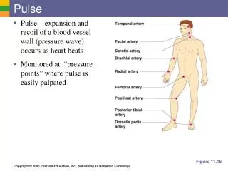
Arterial Pulse. What do u understand by term PULSE ?. The alternate expansion and recoil of elastic arteries after each systole of the left ventricle creating a traveling pressure wave that is called the PULSE. Reading the PULSE Pulses are manually palpated with fingers.

Arterial Pulse
E N D
Presentation Transcript
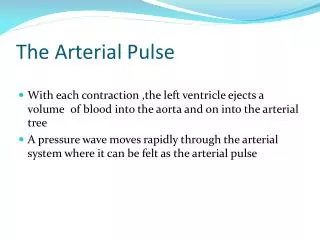
What do u understand by term PULSE? The alternate expansion and recoil of elastic arteries after each systole of the left ventricle creating a traveling pressure wave that is called the PULSE.
Reading the PULSE • Pulses are manually palpated with fingers. • Two or three fingers should be used. • Fingers must be placed near an artery and pressed gently against a firm structure, usually a bone, in order to feel the pulse.
Common pulse sites • Lateral aspect of the lower forearm just proximal to the wrist joint • Feel the bony prominence • Move fingertips medially • Tips of fingers drop into a groove in which lies the artery • Examine the pulse by compressing the artery backwards against the bone, using the finger tips Radial Pulse
Medial aspect of the antecubital fossa at the line of the elbow joint. • The artery is felt by compressing backwards with fingers or thumb through the aponeuosis • Divides just below elbow to form radial and ulnararteries The brachial pulse
1-1.5 cm lateral of the midline in the neck at the upper level of the thyroid cartilage • Readily palpable at anterior border of sternomastoid muscle • May be felt with finger tips or thumb which are used to push posteriorly Carotid pulse
The femoral artery enters the upper leg by passing under the inguinal ligament. • It enters the leg at the mid-inguinal point. • The femoral artery is usually easily palpated and is an important point of access to the arterial system. Femoral artery
The popliteal artery is palpable in the popliteal fossa. • The artery passes through the fossa slightly medially to laterally. • The poplitealartery can be palpated in about the midline of the fossa at the level of the femoral condlyes. • Artery best felt with knee in slight flexion. Popliteal artery
The tibialisposterior artery is found on the medial aspect of the ankle. • It is palpable at a position midway between the prominence of the medial malleolus and the prominence of the calcaneus. Tibialis posterior artery
Dorsalis pedisis a continuation of the tibialis anterior. • Tibialis anterior is often palpable at the ankle joint in a mid-malleolar position, medial to the extensor hallucis longus tendon. Dorsalis pedis artery
Describing the pulse The pulse is described by • Rate • Rhythm • Volume • Synchronous with other pulse or not (Radio-femoral delay). • State of the vessel wall
Rate • The rate of the pulse is recorded in beats per minute. The rate should be counted over a minimum of thirty seconds. • The normal resting pulse rate is 72/min. • Abnormal slow (bradycardia)<60/min • Abnormal fast (tachycardia) >100/min
Rhythm • The rhythm of the pulse is described as regular or irregular. • If irregular the rhythm is described as • regularly irregular (a recurring pattern of irregularity) • irregularly irregular (no discernible pattern to the occurrence Of the irregularity
Volume • The volume of the pulse is a crude indicator of the stroke volume of the heart (the amount of blood ejected by the heart) • It is increased in exercise (full or bounding) and reduced in states of low blood volume (weak or thready)
State of the vessel wall • The normal arterial wall is compressible and has an elastic feel • Diseased arteries may feel inelastic and even hard in cases of calcification
Today’s Lab By the end of this practical the student should be able to: • To locate all peripheral palpable pulses • Examine the radial pulse and comment on pulse rate, rhythm, volume and condition of vessel wall. • Demonstrate the effect of exercise on pulse rate. • Auscultate for heart sounds
Have your partner sit quietly, remaining as relaxed as possible. • Locate the radial pulse • Record the characteristics of the pulse. • Auscultate for the two heart sounds on your partner chest in various areas. • Repeat the procedure and determine the pulse rate and auscultate for heart sounds in each of the following conditions: • Immediately after one minute of exercise • Immediately after 2 minutes of exercise • Immediately after 3 minutes of exercise • 3-5 minutes after exercise has ended.
Switch partners and repeat steps • Tabulate and graph your results and calculate the average pulse of your group
Discussion Questions • Was your pulse higher or lower than your group's average? Suggest reasons for this. • Why can the heart beat be detected as a pulse? • Would wrist or neck pulse be felt first following a heart beat? Why? • If an accident victim had a severed left subclavian artery, which pulse (neck or wrist) would be most affected? Why? • Did you see a difference in your heart rate before and after exercise? Why does this happen? • Did the sound of your heartbeat change after exercise? Describe what differences you heard? • What causes the characteristic heart sounds? • What is pulse deficit and what conditions might cause this?