PULSE
PULSE. K. JAI SHANKAR MD,DM CONSULTANT CARDIOLOGIST INSTITUTE OF CARDIOVASCULAR DISEASES MADRAS MEDICAL MISSION. PULSE. DEFINITION:

PULSE
E N D
Presentation Transcript
PULSE K. JAI SHANKAR MD,DM CONSULTANT CARDIOLOGIST INSTITUTE OF CARDIOVASCULAR DISEASES MADRAS MEDICAL MISSION
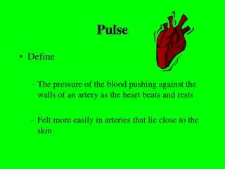
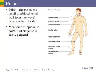
PULSE DEFINITION: • Pulse is the palpability over peripheral arteries, a pulse wave which is a transmitted wave from the root of aorta along the vessel wall traveling 10 times faster than blood. • Blood travels at speed of - .5 mt/sec. • Pulse travels at speed of - 5 mt/sec.
PULSE WAVE • The arterial pulse reflects the performance of LV • “Mirror of the heart” • It is propagated by incompressible blood both forwards and laterally. The lateral movement distends the arterial wall and is felt as pulse.
PULSE - HISTORY HIPPOCRATES – 4TH CENTURY BC Thought that arteries are air ducts GALEN Arteries contain blood & not air. HEROPHILUS Recognized that arterial pulses & cardiac pulses were synchronous.
PULSE - HISTORY Nei Ching Su Weri – The yellow emperors book of medicine. The oldest book of medicine still existing. It quotes that chief means of diagnosis than was pulse. • It was palpated for hours in a dozen sites • It was noted whether strong or weak regular or irregular • At that time as watches were not invented pulse was timed by the physicians respiratory excursions.
Determinents of Arterial pulse Left Ventricle: Stroke volume LV contractility Velocity of LV ejection Aortic Valve : Normal Stenosis Regurgitation Both stenosis and regurgitation Arterial system: Compliance or distensibility Peripheral vascular resistance Aortic run off
BLOOD FLOW LV pressure when it rises above aortic pressure becomes driving force for movement of blood into aorta Driving force is dependent on 1) Contractility 2) Size & shape of LV 3) Heart rate. This driving force is opposed by several forces that impede the flow 1) Resistance2) Inertia3) Compliance
SYSTOLIC UPSTROKE TIME Onset of pulse wave to its peak Normal range = 90-160 ms Brachial artery = 120 ms • Acceleration time in Echo
PULSE WAVE COMPONENTS • Percussion wave is impulse generated by LV ejection • Tidal wave is percussion wave reflected from upper part of the body • Dicrotic wave is reflected from lower part of the body often recorded but not palpable • Anacrotic notch occurs towards the end of rapid ejection phase just before max pressure is reached • Incisura Occurs in Isovolumic relaxation phase prior to aortic valve closure. • Upstroke comes with S1 • Peak is reached well before S2
CENTRAL PULSE • The central pulse begins with AV opening and onset of LV ejection • The rapid rising portion of the arterial pressure curve is termed anacrotic limb (Greek – upbeat) • An anacrotic notch is frequently recorded on the ascending limb towards the end of rapid ejection phase. • Peak Aortic flow velocity occurs slightly earlier than the peak pressure. • The Pulse shows 2 systolic waves “Percussion wave” and “Tidal wave”
CENTRAL PULSE • The descending limb of the carotid arterial pulse is less steep than the ascending limb • The descending limb is interrupted by a incisura a sharp downward deflection in end systole related to isovolumic relaxation phase • The subsequent small positive “dicrotic wave” is attributed to 1) Elastic recoil of aorta and AV 2) Reflected waves from most distal arteries.
ALTERATIONS IN CENTRAL PULSE PERIPHERALLY • Upstroke becomes steeper • Systolic peak becomes higher • Anacrotic notch disappears • Systolic upstroke time becomes shorter (120msec)
ALTERATIONS IN CENTRAL PULSE PERIPHERALLY • Systolic ejection time becomes more (320msec) • The dicrotic notch occurs much later • Systolic pressure increases • Diastolic pressure & mean pressure decreases
CAUSES FOR CHANGE IN CENTRAL PULSE CONTOUR WHEN TRANSMITTED PERIPHERALLY 1)Distortion & damping of pulse wave components 2) Different rates of transmission of various components 3) Differences in distensibility & caliber of arteries 4) Changes in the vessel wall due to age & or disease
CHANGES IN PULSE WITH AGING 1) Increase in the height of tidal wave 2) Increase in the height of the incisura 3) Systolic upstroke time is longer 4) Amplitude & duration of dicrotic wave decreases Normally PW is taller than TW and TW is not palpable. In old age, diabetes & arteriosclerosis TW is taller and this is clinically appreciated as the pulse reaching a peak in late systole.
PERIPHERAL ACCESSIBLE ARTERIES 1) Head & Neck 1) Superficial Temporal 2) Carotids 3) Subclavian 2) Upper Limb 4) Axillary 5) Brachial 6) Radial 3) Abdomen 7) Abdominal aorta 4) Lower Limb 8) Femoral 9) Popliteal 10) Posterior Tibial 11) Dorsalis Pedal
Localization of arteries • The CCA terminates at C4 level at upper border of thyroid cartilage • The ECA is palpated medial to the sternocleidomastoid above upper border of the thyroid cartilage • The ICA is palpated placing a hand in the mouth and palpating the tonsillar fauces. • The subclavian artery is felt in the posterior triangle. With the shoulder depressed, pressure is exerted down back and medially in the angle between sternocleidomastoid and clavicle.
Localization of arteries • Brachial-Palpation of the right brachial pulse is accomplished with the thumb of the examiners right hand as the patients arm lies supinated at his or her side • Axillary- compression against the humerus.
RADIAL For radial pulse palpation the pts hand should be supinated & comfortably supported. The examiners thumb or tip of a single finger preferably the index is applied to the pulse. In infants palpation of radial pulse has inherent limitations 1) Radial artery is very small 2) Padding of subcutaneous fat is more.
EVALUATION OF ARTERIAL PULSE 1) Rate& rhythm 2) Volume &tension 3) Character 4) Vessel wall 5) Peripheral pulses Grade the palpability Brachio or radio- femoral and brachio-brachial delay Bruit Palpation of abdominal artery Ocular fundi Allen’s test
GRADING OF PULSES GRADE 0 -absent pulse + - feeble ++ - palpable but diminished compared to other side +++ - normal ++++ - high volume or bounding pulse
ABNORMAL PULSES 1) Pulsus Parvus 2) Pulsus Tardus 3) Hypokinetic Pulse 4) Hyperkinetic Pulse ( Bounding) 5) Brisk or Jerky Pulse 6) Water Hammer Pulse 7) Collapsing Pulse 8) Corrigans Pulse 9) Anacrotic Pulse 10) Bisferrians Pulse 11) Dicrotic Pulse 12) Pulsus Paradoxsus 13) Pulsus Alternans 14) Pulsus Bigeminny
PULSUS PARVUS A slow rising pulse Low volume pulse Best appreciated in carotids Seen in severe AS and severe heart failure.
PULSUS TARDUS( Anacrotic pulse) Late peaking Peak is delayed and nearer to S2 Best appreciated by simultaneous auscultation of the heart and palpation of carotid pulse Seen in all forms of fixed obstruction to the LVOT
ANACROTIC PULSE Pulsus parvus et tardus with accentuation of the anacrotic notch and a small volume pulse. Characterized by- 1)Slow upstroke 2)Delayed peak 3)Small volume
CHARACTERISTICS OF ANACROTIC PULSE 1)Pulsusparvus 2)Pulsustardus 3)Small volume 4)Prominent anacrotic notch which appears earlier 5)Dicrotic notch disappears It is well felt in the carotids Earlier the anacrotic notch severe the stenosis→ correlates with a gradient of 70 mmHg
Normal arterial pulse with AS • Mild AS • Associated AR • HOCM • Supravalvular AS, CoA • In children and elderly
HYPOKINETIC PULSE Small or diminished pulse 1) Low CO 2) LV Dysfunction 3) CCF 4) Hypotension 5) LVOT Obstruction In Hypokinetic pulse Normal upstroke indicates decreased SV Slow uprise indicates LVOT obstruction
HYPERKINETIC PULSE • Anxiety 2) Anaemia 3) Thyrotoxicosis 4) Exercise 5) Hot humid environment 6) Alcohol intake 7) Cigarette smoking 8) SHT with Atherosclerosis 9) Isolated Systolic HT
HYPERKINETIC PULSE Hyperkinetic pulse has a larger than normal amplitude and results from 1) Increased LV ejection velocity 2) Increased Stroke volume 3) Increased arterial pressure.
JERKY PULSE Jerky pulse is a pulse with a brisk or sharp upstroke that literally taps against the palpating fingers. The pulse volume is not increased Rapid upstroke / Normal downstroke / Normal volume Seen typically in HCM
COLLAPSING OR WATER HAMMER PULSE Thomas Watson(1844) coined the term after victorian toy. The collapsing pulse is due to : i) Diastolic run off into the LV ii) Reflex vasodilatation mediated by carotid baroreceptors secondary to large stroke volume iii) Rapid run off from the periphery due to decreased systemic vascular resistance. Best appreciated at the radial pulse with the palmer side of the examiner’s hand and with the patient’s arm suddenly elevated above the shoulder. This may be related to the artery becoming more in the line with the central aorta, allowing direct systolic ejection and diastolic backward flow.
COLLAPSING PULSE With aortic run off: AR, PDA, AP window, RSOV into right side and AV fistula. Cyanotic CHD : • Truncus arteriosus with truncal run off in to PA or truncal insufficiency, • Pulmonary atresia with AP collaterals, • TOF with AP collaterals/associated PDA/ associated AR / after BT shunt. Hyperkinetic states Pregnancy, Anemia, thyrotoxicosis, Beriberi, Fever, Paget’s disease of Bone Normal Volume Collapsing Pulse 1) MR 2) VSD
PERIPHERAL SIGNS OF AR HEAD & NECK 1) De Mussets sign Head bobbing 2) Light House Sign Alt flushing & blanching of face 3) Landolfis sign Alteration in pupillary size with cardiac cycle 3) Quinckies sign Capillary pulsation over lips 4) Mullers sign Uvula pulsation 5) Carotid shudder Thrill over carotid during upstroke 6) Corrigans Pulse Visible carotid pulse of AR 7) Julians sign Pulsation of retinal vessels. 8) Minervini’s sign Strong lingual pulsations. Tongue depressor moves up and down when tongue is depressed. 9) Logue’s signPulsation of sternoclavicular junction when AR is associated with aortic dissection.
PERIPHERAL SIGNS OF AR LIMBS 10) Bisferiens Pulse Double peaked Pulse 11) Locomotor Brachi Dancing Brachialis 12) Hills sign LL SBP > 20 mm than UL Mild 20-40 mmhg Moderate 40-60 mmhg severe >60mmhg 13) Pistol shot Femoralis Systolic sounds over FA 14) Traubes sign Systolic & Diastolic sounds 15) Durozies murmur. Distal occlusion diastolic murmur Proximal occlusion systolic murmur 16)Palfrey’s sign Pistol shot sound over radial artery
PERIPHERAL SIGNS OF AR ABDOMEN 17) Rosenbachs sign - Liver Pulsation 18) Gerhardts sign - Splenic Pulsation 19) Dennison’s sign - Presence of pulsations in cervix
Bisferiens pulse Normally percussion wave is felt but not the tidal wave. In all the conditions where percussion wave is prominent, tidal wave also becomes prominent. Mechanism: In combined AS and AR, the stenotic component permits a jet, & lateral to the jet there is a fall in pressure( Bernoulli Phenomenon), this results in a dip or inward movement in the pulse with secondary outward movement in a pulse or tidal wave.
Bisferiens pulse Normally both waves are prominent in patients with severe AR. In HOCM, the initial part of left ventricular ejection is rapid, resulting in rapid upstroke. As obstruction to the outflow starts later in the systole, due to SAM, a sudden interruption to left ventricular ejection occurs resulting in a dip in the pressure pulse followed by the slow rising pulse wave, which is characteristic of HOCM ( spike and dome pattern). The percussion wave is more prominent than tidal wave in HOCM. Seen in Severe AR,AS with AR,HOCM,hyperkinetic circulatory state,after exercise
DICROTIC PULSE Dicrotic pulse has an accentuated dicrotic wave and hence is a twice beating pulse, one in systole and one in diastole. Requirements : 1) Hypotension 2) Reduced Peripheral Vascular Resistance When the reflection wave travels rapidly and meets the original wave well in advance, it is lost in it. In rigid and nondistensible arterial system, as in SHT, dicrotic pulse in never present. It is differentiated from the bisferiens pulse by the simultaneous auscultation of the heart sounds.
DICROTIC PULSE It is more noticeable in the beat following a PVC. It is better appreciated during inspiration or inhalation of amyl nitrite. IABP-augmented wave due to diastolic flow occlusion in descending aorta Rarely present when BP > 130 mmHg and in patients beyond 50 years of age.
DICROTIC PULSE 1) Healthy young adults 2) Fever 3) Hypovolemic shock 4) CCF 5) Cardiac tamponade 6) Sepsis 7) Post AVR 8) IABP
TWICE BEATING PULSE Anacrotic, Bisferiens ,Dicrotic Differentiation: The double peaking occurs A) On the upstroke in Anacrotic;late peaking B) On the peak in Bisferiens- Both in Systole;rapid rising C) On the downstroke in Dicrotic ; normal rising One in Systole & One in Diastole
PULSUS PARADOXUS Paradox about the pulse is absence of pulse during inspiration but presence of heart sounds & was coined by Adolph Kussmaul in 1873. Suspected if the pulse varies with inspiration in all accessible arteries. MISNOMER- the term paradoxus is that normally there is a fall in BP during inspiration (4-6mm/hg) which in PP is exaggerated (>10mm/hg)