Structure-function relationship: Fibrous proteins
Structure-function relationship: Fibrous proteins. Lecture 4 Dr. Mamoun Ahram Summer, 2014. Biological Functions of Proteins. Enzymes--catalysts for reactions Transport molecules--hemoglobin; lipoproteins, channel proteins Contractile/motion--myosin; actin

Structure-function relationship: Fibrous proteins
E N D
Presentation Transcript
Structure-function relationship:Fibrous proteins Lecture 4 Dr. Mamoun Ahram Summer, 2014
Biological Functions of Proteins • Enzymes--catalysts for reactions • Transport molecules--hemoglobin; lipoproteins, channel proteins • Contractile/motion--myosin; actin • Structural--collagen; keratin, actin • Defense--antibodies • Signaling—hormones, receptors • Toxins--diphtheria; enterotoxins
Types of proteins • Proteins can be divided into two groups according to structure: • fibrous (fiber-like with a uniform secondary-structure only) • globular (globe-like with three-dimensional compact structures) • Examples • Fibrous proteins: collagens, elastins, and keratins • Globular proteins: myoglobin, hemoglobin, and immunoglobulin
The extracellular matrix • The extracellular space is largely filled by an intricate network of macromolecules including proteins and polysaccharides that assemble into an organized meshwork in close association with cell surface.
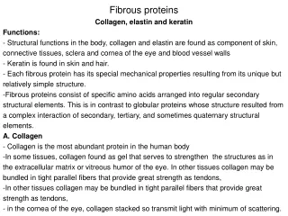
Collagens and their properties • The collagens are a family of fibrous proteins with 25 different types found in all multicellular animals. • They are the most abundant proteins in mammals, constituting 25% of the total protein mass in these animals. • Collagen molecules are named as type I collagen, type II collagen, type III collagen, and so on. • The main function of collagen molecules is to provide structural support to tissues. • Hence, the primary feature of a typical collagen molecule is its stiffness.
Structure • It is a left-handed, triple-stranded, helical protein, in which three collagen polypeptide chains, called chains, are wound around one another in a ropelike superhelix. • This basic unit of collagen is called tropocollagen. • Compared to the a-helix, the collagen helix is much more extended with 3.3 residues per turn.
Composition of collagens • Collagens are rich in glycine (33%) and proline (13%). • It is also unusual in containing 4-hydroxyproline (9%) and hydroxylysine • Every third residue is glycine, which, with the preceding residue being proline or hydroxyproline in a repetitive fashion as follows: • Gly-pro-Y • Gly-X-hydroxyproline
Functional purpose of amino acids • Glycine allows the three helical a chains to pack tightly together to form the final collagen superhelix. • Proline creates the kinks and stabilizes the helical conformation in each a chain.
Hydroxylysine • Hydroxylysineserves as attachment sites of polysaccharides making collagen a glycoprotein
Oxidation of lysine • Some of the lysine side chains are oxidized to aldehyde derivatives. • Covalent aldol cross-links form between hydroxylysine residues and lysine or another oxidized lysine.
Function of cross-linking • These cross-links stabilize the side-by-side packing of collagen molecules and generate a strong fibril • If cross-linking is inhibited, the tensile strength of the fibrils is drastically reduced; collagenous tissues become fragile, and structures such as skin, tendons, and blood vessels tend to tear. • The amount of cross-linking in a tissue increases with age. That is why meat from older animals is tougher than meat from younger animals.
Formation of collagen fibers • Following cellular release of protocollagen, 5 of them polymerize into a microfibril, which are connected with each other via aldehyde links. • Microfibrils align with each other forming larger collagen fibrils, which are strengthened by the formation of covalent cross-links between lysine residues. • Microfibrils assemble into collagen fibers.
Purpose of hydroxyproline • Normal collagen is stable even at 40 °C. • Without hydrogen bonds between hydroxyproline residues, the collagen helix is unstable and loses most of its helical content at temperatures above 20 °C
Scurvy • Scurvy is a disease is caused by a dietary deficiency of ascorbic acid (vitamin C). • Deficiency of vitamin C prevents proline hydroxylation. • The defective pro-α chains fail to form a stable triple helix and are immediately degraded within the cell. • Blood vessels become extremely fragile and teeth become loose in their sockets.
Resilience vs. flexibility • Many tissues, such as skin, blood vessels, and lungs, need to be both strong and elastic in order to function. • A network of elastic fibers in the extracellular matrix of these tissues gives them the required resilience so that they can recoil after transient stretch. • Long, inelastic collagen fibrils are interwoven with the elastic fibers to limit the extent of stretching and prevent the tissue from tearing
Elastin • The main component of elastic fibers is elastin, which is a highly hydrophobic protein and is rich in proline and glycine. • It contains some hydroxyproline but no hydroxylysine. • It is not glycosylated • The primary component, tropoelastin molecules, is cross-linked between lysines to one another.
Elastin structure • The elastin protein is composed largely of two types of short segments that alternate along the polypeptide chain: • hydrophobic segments, which are responsible for the elastic properties of the molecule; and • alanine- and lysine-rich -helical segments, which form cross-links between adjacent molecules
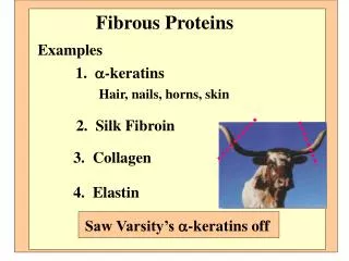
Keratins • Two important classes of proteins that have similar amino acid sequences and biological function are called -and -keratins, which as members of a broad group of intermediate filament proteins. • -keratin is the major proteins of hair and fingernails as well as animal skin. • -keratin has an unusually high content of cysteine.
-keratins structure (hair vs. fingernails) • Two helical -keratin molecules interwine. • Two dimers twist together to form a 4-molecule protofibril. • Eight protofibrils combine to make the microfibril. • Hundreds of microfibrils are cemented into a macrofibril. • -keratin is hardened by the introduction of disulfide cross-links (fingernails).
How is a perm done? A basic reducing substance (usually ammonium thioglycolate) is added to reduce and rupture some of the disulfide cross-links Temporary Wave Permanent wave When the hair gets wet, water molecules intrude into the keratin strands disrupting some of the hydrogen bonds, which help to keep the alpha-helices aligned. When hair is dried up, the hair strands are able for a short time to maintain the new curl in the hair. The hair is put on rollers or curlers. The alpha-helices can shift positions. An oxidizing agent, usually a dilute solution of hydrogen peroxide, is added to reform the disulfide bonds in their new positions. The permanent will hold these new disulfide bond positions until the hair grows out.