Download
1 / 27
270 likes | 395 Vues
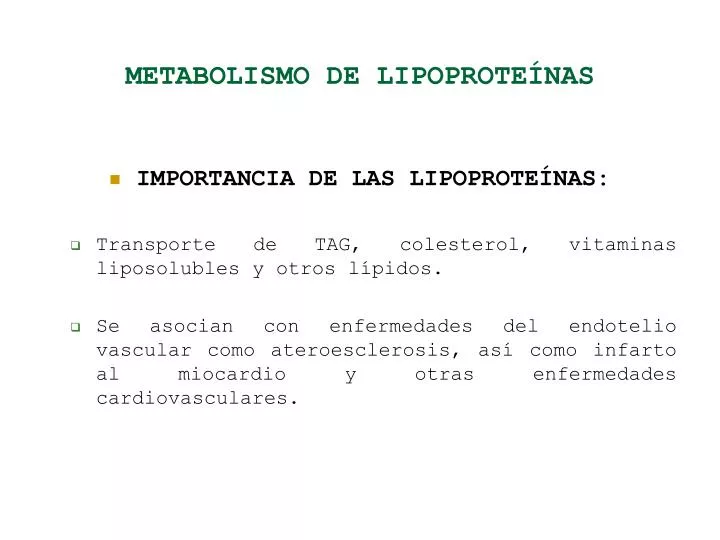
METABOLISMO DE LIPOPROTEÍNAS. IMPORTANCIA DE LAS LIPOPROTEÍNAS: Transporte de TAG, colesterol, vitaminas liposolubles y otros lípidos. Se asocian con enfermedades del endotelio vascular como ateroesclerosis, así como infarto al miocardio y otras enfermedades cardiovasculares.

E N D
METABOLISMO DE LIPOPROTEÍNAS • IMPORTANCIA DE LAS LIPOPROTEÍNAS: • Transporte de TAG, colesterol, vitaminas liposolubles y otros lípidos. • Se asocian con enfermedades del endotelio vascular como ateroesclerosis, así como infarto al miocardio y otras enfermedades cardiovasculares.
ESTRUCTURA DE LAS LIPOPROTEÍNAS • Núcleo de ésteres de colesterol y TAG. • Capa externa con fosfolípidos, colesterol libre y apoproteínas.
Figure 17.2 The lipoprotein particle. The external monolayer of a lipoprotein particle contains free cholesterol, phospholipids, and apoproteins. Cholesterol esters and triacylglycerols locate in the particle core.
ESTRUCTURA DE LAS LIPOPROTEÍNAS • Clasificadas según su densidad: • Quilomicrones: TG, B48, 75-1200 μm diámetro. • VLDL: TG, B100, 30-80. • IDL: TG y CHOL, B100, 25-35. • LDL: CHOL, B100, 18-25. • HDL: Proteína, AI, AII, 5-12.
ESTRUCTURA DE LAS LIPOPROTEÍNAS • Apoproteínas: • Interactúan con receptores. • Activan o inhiben enzimas de metabolismo de lipoproteínas. • ApoA: en HDL. • ApoB: metabolismo de quilomicrones y LDL. • ApoE: unión a receptores. • ApoC: activador e inhibidor enzimático.
ESTRUCTURA DE LAS LIPOPROTEÍNAS • Lipoproteína (a): • Partícula LDL (con apoB100), mediante puentes disulfuro se une a: • Apo (a). • Sintetizada en el hígado, homologa a plasminógeno.
Figure 17.3 Schematic structure of lipoprotein (a). Lipoprotein (a) is essentially an LDL particle, with apo(a) is linked to apoB through a disulfide bridge. Apo(a) is a large molecule containing a number of repeat units (kringles). Kringles have structure similar to plasminogen.
FUNCIONES DE LAS LIPOPROTEÍNAS • Quilomicrones: • Transporte de TAG de intestino a tejidos periféricos. • Lipoproteín lipasa hidroliza quilomicrones en el hígado. • Productos de hidrólisis son captados por receptor de LDL al que se une la apoB48 del quilomicrón.
FUNCIONES DE LAS LIPOPROTEÍNAS • VLDL: • Transporte de TAG del hígado a tejidos periféricos. • Lipoproteín lipasa hidroliza VLDL que se convierte en IDL o LDL. • ApoE (en IDL) o apoB100 (en LDL) se unen a receptores hepáticos.
FUNCIONES DE LAS LIPOPROTEÍNAS • LDL: • Transporte de CHOL de hígado a periferia. • HDL: • Transporte de CHOL de la periferia al hígado.
ENZIMAS Y PROTEÍNAS ACARREADORAS • Lipasa de lipoproteínas: • Hidroliza TAG de lipoproteínas en endotelio vascular. • Actúa sobre quilomicrones y VLDL, libera glicerol y AGL. • Triacilglicerol lipasa hepática: • Hidroliza TAG en membrana plasmática hepática. • Actúa sobre partículas digeridas parcialmente por la lipasa de lipoproteínas (IDL, LDL).
ENZIMAS Y PROTEÍNAS ACARREADORAS • Lecitín-colesterol aciltransferasa: • Enzima asociada con HDL. • Esterifica CHOL tomado por HDL.
RECEPTORES DE LIPOPROTEÍNAS • Principal receptor: receptor de apo B/E (conocido como receptor de LDL). • La expresión del gene del receptor está regulada por la concentración intracelular de colesterol.
Figure 17.4 LDL and scavenger receptors. While the LDL (apoB/E) receptor mediates the uptake of intact LDL, the scavenger receptor internalizes modified LDL. Both receptors span the cell membranes. The expression of LDL receptor is regulated by the intracellular cholesterol concentration, while the scavenger receptor remains unregulated. The scavenger receptor type A, which is illustrated here, is present on macrophages and has a collagen-like structure. Scavenger receptor type BI participates in HDL-metabolism.
RECEPTORES DE LIPOPROTEÍNAS • Receptores de amplia especificidad: “scavenger”. • En células fagocíticas como los macrófagos. • No regulados por mecanismos de retrocontrol.
Figure 17.5 (B) The calculation of LDL-cholesterol concentration in plasma. LDL-cholesterol can be calculated from the value of total cholesterol, triglycerides, and HDL-cholesterol using the Friedewald formula.
Figure 17.6 Lipoprotein metabolism: the fuel transport pathway and the overflow pathway. The fuel transport pathway: chylomicrons transport triglyceride to the periphery, and their remnants are metabolized in the liver. VLDL transport fuel from the liver to peripheral tissues, and the remnants also return to the liver. Part of VLDL remnants and IDL are further converted into LDL, which enter the overflow pathway. The overflow pathway: LDL travel in blood from the liver through the peripheral tissues and back to the liver. On its way it may enter the arterial wall. The amount of lipids deposited in the arteries is proportional to their plasma concentration. Cholesterol is removed from cells and transported back to the liver by HDL particles. LPL: lipoprotein lipase, LRP: LDL-receptor-related protein.
Figure 17.7 Reverse cholesterol transport. HDL are assembled in the liver and intestine as discoidal particles. They acquire cholesterol from cell membranes aided by cholesterol efflux regulatory protein (CERP). LCAT associated with HDL esterifies the acquired cholesterol. The formed cholesteryl esters move to the inside of the particle, and the particle becomes spherical. HDL exchanges apoproteins and cholesteryl esters with triacylglycerol-rich lipoproteins. This is facilitated by cholesterol ester transfer protein (CETP). HDL acquire triacylglycerols in exchange for cholesteryl esters. This increases their size further. However, when cholesterol transfer to the liver mediated by the scavenger receptor BI is completed, the HDL size decreases again: some of the redundant material is used to construct apoAI-rich, lipid-poor particles (pre-beta HDL). These re-enter the cholesterol removal cycle.
Figure 17.8 Atherogenesis: the process. Atherogenesis is driven by signals mediated by cytokines and growth factors generated by all the major types of cells participating in the process: endothelial cells, macrophages, T lymphocytes and vascular smooth muscle cells (VSMC). There are multiple activation paths: for instance, the expression of MCP-1 and VCAM-1 may be stimulated by signals generated macrophages as well as by the oxidized LDL. VSMC may be stimulated by the dysfunctional endothelial cells, by macrophages, and by T lymphocytes (note also the autocrine activation). Note that a hormone, angiotensin II also participates in these processes. MCP-1: monocyte chemoattractant protein 1, VCAM-1: vascular cell adhesion molecule 1, ICAM-1: intracellular cell adhesion molecule 1, TNFβ: tumor necrosis factor beta, TNFα: tumor necrosis factor alpha, IFNγ: interferon gamma, NO: nitric oxide, PDGF: platelet-derived growth factor, bFGF: basic fibroblast growth factor, IGF-1: insulin-like growth factor 1, EGF: epidermal growth factor, TGFβ: transforming growth factor beta, IL-1: interleukin 1.
Figure 17.9 Atherogenesis: role of growth factors and cytokines. Atherogenesis involves endothelial dysfunction, arterial deposition of lipids, inflammatory reaction, and the migration and proliferation of the arterial smooth muscle cells. Note the key role of lipid oxidation in the formation of lipid-laden cells and the lipid center of the atherosclerotic plaque. The sequence of events, and their control by cytokines and growth factors are described in the text. (Compare Fig. 17.8.)
Figure 17.10 Atherosclerotic plaque. The lipid center and fibrous cap are the main parts of a mature atherosclerotic plaque which emerges from the structurally changed vascular wall. The so-called vulnerable plaque ruptures easily. This figure illustrates areas vulnerable to breakage and shows the obstructing thrombus formed at the rupture site.
Figure 17.11 Plasma cholesterol and coronary mortality in men. Total cholesterol concentration in plasma in relation to the number of deaths from heart disease in a population. Note: converting plasma lipid values into SI units: cholesterol: to convert mg/dL into mmol/L, mutliply by 0.02586; triglycerides: to convert mg/dL into mmol/L, multiply by 0.01129. The data are from the Multiple Risk Factor Intervention Trial (MRFIT). Stammler et al. JAMA 1986;256:2823.
Figure 17.12 The interpretation of cholesterol, triglyceride, and HDL-cholesterol measurements. To convert plasma lipid values into conventional units: total cholesterol, LDL-cholesterol, and HDL-cholesterol: to convert mmol/L into mg/dL: multiply by 38.67; triglycerides: to convert mmol/L into mg/dL, multiply by 87.5. CHD, coronary heart disease.