Download
1 / 55
550 likes | 900 Vues
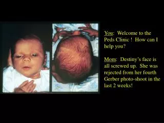
You : Welcome to the Peds Clinic ! How can I help you? Mom : Destiny’s face is all screwed up. She was rejected from her fourth Gerber photo-shoot in the last 2 weeks! . Normal Infant Skull. Expansile. Flexible enough to get through vagina Molding

E N D
You: Welcome to the Peds Clinic ! How can I help you? Mom: Destiny’s face is all screwed up. She was rejected from her fourth Gerber photo-shoot in the last 2 weeks!
Normal Infant Skull Expansile • Flexible enough to get through vagina • Molding • Expansile enough to accommodate rapid brain growth Flexible
Suture Growth • Sutures allow growth perpendicular to them • Growth at suture lines related to brain growth
Ossification By Age 8 Bone Union By Age 20 Suture Closure
Craniosynostosis: Early Fusion of a Suture
Sagittal Synostosis “Boat-Head” (Scaphocephaly)
Coronal Synostosis “Bent-Head” (Plagiocepahly)
Metopic Synostosis “Triangle-Head” (Trigonocephaly)
Lamboid Synostosis MATTRESS “Slant-Head” (Occipital Plagiocephaly)
Clinical Exam • OFC • Head shape (from above, side) • Ear and facial symmetry • Palpate suture lines & fontanelles • Look for ridging • Look for associated anomalies • Skull X-ray or CT
Craniosynostosis Secondary Primary Microcephaly Prematurity VP Shunting Positioning Isolated Abnormal Suture Syndromic
Prematurity • Deformational Scaphocephaly • Impaired mobility & prolonged positioning • Persists until adulthood • Prevention: • Donut-shaped head supports • waterbed mattresses • Does not warrant intervention
VP Shunting • Scaphocephaly • Chronic hydrocephalus thickens the skull • Once decompression with shunt, the suture fuses • Surgery Indications: • OFC > 50 cm (4-5+ STDs) • When VPS performed during when VLBW
Microcephaly • Surgical correction not indicated • Abnormal OFC • in primary craniosynostosis, OFC remains normal yet oddly shaped • Rare cases of multisutural craniosynostosis restricting head growth, but manifests with increased ICP
PositionalDeformation • Most common cause • Usually forehead asymmetry • Sometimes associated with torticollis • Usually acts on coronal or lamboidal suture • 40% of newborns
An Epidemic of Lamboidal Plagiocephaly • 1992: Back to Sleep • Campaign • 1996: Tertiary Care • Centers report rise in • lamboidal plagiocephaly • from 3% to 20%
Sorting out the “Epidemic” • 102 Patients with occipital plagiocephaly over 4 year period • Only 4 (3%) had true lamboidal synostosis • The rest were deformational • Only 3 were progressive (required surgery) • Other responded to positioning or helmets
Syndromic Craniosynostosis • 10-20 % of cases • Autosomal Dominant • Linked to Chromosome 10q • Multi-sutural, complex cases If a suture is fused, check hands, feet, big toe and thumb
Distinguishing Clinical Features in the Craniosynostosis Syndromes
Crouzon’s • Normal intellect • Normal extremities • 5 % have acanthosis nigricans • 30 % have progressive hydrocephalus
Apert’s“Crouzon’s with Hand Involvement” • Varying intellect (50 % with MR) • Mitten Glove Syndactyly • Cervical vertebral anomalies • Rare hydrocephalus
True Craniosynostosis & Surgery • Single Suture Synostosis: Confirm by exam and skull x-rays • Complex cases: CT or 3D CT • X-Ray: Fused sutures have a broad ridge of overgrowth of solid bone along a previous suture, or suture is completely obliterated • Ridge is especially characteristic of fused sagittal suture
Management • Surgery vs. Conservative Management
The Decision to Operate • Raised ICP in 1/3 of cases, but no neuro impairment • Cosmetic considerations usually most important • affects peer acceptance, parent-child bonding, self-image and coping
Imaging • Skull X-ray • CT • 3-D CT
Surgery • If not part of syndrome, the earlier the operation the better • At the latest 6-12 months (by 12 months, skull is 85% of adult size) • For coronal suture, operate before 2 months because of facial symmetry and visual system development • Procedure depends on continuing skull growth • Hospitalization for 3-10 days
Surgery • Syndromic cases may need special airway support • Blood loss significant due to scalp vascularity • transfusion rates 20-500 % of infant estimated blood volume • PICU stay (facial edema) • Results on xray within several days
Metopic Synostosis
Surgery • Unilateral coronal suture: difficult. Orbital relocation as well. • Syndromic or multi-suture cases: staged repairs.