The Abdomen
The Abdomen. Surface Anatomy, Vessels, Muscles, and Peritoneum. Hastaneciyiz.blogspot.com. Ventral body cavity Thoracic Abdominopelvic Abdominopelvic Abdominal Liver Stomach Kidneys Pelvic cavity Bladder Some reproductive organs Rectum. Abdominopelvic Cavity.

The Abdomen
E N D
Presentation Transcript
The Abdomen Surface Anatomy, Vessels, Muscles, and Peritoneum Hastaneciyiz.blogspot.com
Ventral body cavity Thoracic Abdominopelvic Abdominopelvic Abdominal Liver Stomach Kidneys Pelvic cavity Bladder Some reproductive organs Rectum Abdominopelvic Cavity
Surrounded by the abdominal walls and pelvic girdle The two cavities are continuous Most organs surrounded by a peritoneal cavity Visceral peritoneum Serous peritoneum Peritoneal cavity Abdominopelvic Cavity pg 242
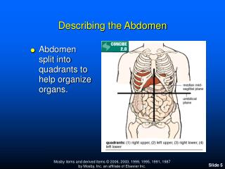
9 regions 4 quadrants Draw “line” through navel Right upper quadrant Left upper quadrant Left lower quadrant Right lower quadrant Abdominal Quadrants pg 242
Anterior abdominal wall extends from costal margin to inferior boundaries: Iliac crest Anterior superior iliac spine Inguinal ligament Pubic crest Superior boundary Diaphragm Central landmark Umbilicus Linea alba (white line) Tendinous line Extends from xiphoid process to pubic symphysis Surface Anatomy pg 345
Function: Help contain abdominal organs Move trunk Forced breathing Increase intra-abdominal pressure Abdominal wall Anterior (4) Innervated by intercostal nerves Continuous with layers of intercostal muscles Fibers of layers run in different directions for strength Ends in aponeurosis which contains rectus abdominis muscle Posterior (3) Muscles pg 250
Rectus Abdominis Origin Pubic crest, symphysis Insertion Xiphoid process, costal cartilages of ribs 5-7 Function Flex, rotate trunk, fix and depress ribs, stabilize pelvis, compress abdomen Internal oblique Origin Lumbar fascia, iliac crest, inguinal ligament Insertion Linea alba, pubic crest, last 3-4 ribs, costal margin Function Same for external obliques Anterior Abdominal Wall Muscles pg 250, 251
External oblique Origin Lower 8 ribs Insertion Aponeurosis to linea alba, pubic and iliac crest Function Flex trunk, compress abdominal wall (together), Rotate trunk (separate sides) Transversus abdominis Origin Inguinal ligament, lumbar fascia, cartilage of last 6 ribs, iliac crest Insertion Linea alba, pubic crest Function Compress abdominal contents Anterior Abdominal Wall pg 249
Iliopsoas Psoas major Origin Lumbar vertebrae, T12 Insertion Lesser trochanter of femur via iliopsoas tendon Function Thigh flexion, trunk flexion, lateral flexion Innervation Ventral rami L1-L3 Iliacus Origin Iliac fossa, ala of sacrum Insertion Lesser trochanter of femur via iliopsoas tendon Function Thigh flexion, trunk flexion Innervation Femoral nerve (L2 and L3) Psoas minor – variable (40-60% do not have) Posterior Abdominal Wall pg 316
Quadratus lumborum Origin Iliac crest and lumbar fascia Insertion Transverse process of upper lumbar vertebrae, lower margin of rib 12 Function Flex vertebral column, maintains upright posture, assists in inspiration Innervation: T12 and upper lumbar spinal nerves (ventral rami) Posterior Abdominal Wall pg 316
Peritoneum • Mesenteries • Double layer of peritoneum (2 serous membranes fused together) • Extend to the digestive organs from the body wall • Function: • Hold organs in place • Sites of fat storage • Provide a route for vessels and nerves • Dorsal mesenteries: • Lesser omentum and Falciform ligament • Ventral mesenteries: • Greater omentum, Transverse mesocolon, Mesentary, and Sigmoid mesocolon
Dorsal Mesenteries pg 291 pg 269
Ventral Mesenteries pg 271 pg 269
Peritoneum • Peritoneal • Remains surrounded by peritoneal cavity • Liver, stomach, ileum and jejunum • Retroperitoneal • Some organs lay behind/outside peritoneum • Primarily retroperitoneal • Organs NEVER within the cavity • Kidneys, bladder, ureter • Secondarily retroperitoneal • Organs once suspended within the abdominal cavity by mesentery • Migrate posterior to the peritoneum during the course of embryogenesis to become retroperitoneal • Lack mesenteries • Duodenum, ascending and descending colon, rectum, pancreas
Organs of the Abdomen Urinary and Digestive Systems
Kidney (2) Purify blood Ureter (2) Drains urine from kidney to bladder Urinary Bladder Stores urine Urethra Drains urine from bladder to outside body Urinary System pg 314
Kidneys • Filter waste from blood • Water, toxins, urea, uric acid, creatinine, metabolic waste, ions • Excretion of waste • Homeostasis • Acid-base balance • Blood pressure • Plasma volume
Lie in retroperitoneal, superior lumbar region Extend from T11 or T12 to L3 Laterally convex, medially concave Hilus Where blood vessels, ureters, and nerves enter and leave kidney Adrenal gland On superior portion Kidneys: Gross Anatomy pg 325
Separated into lobes Blood supply Renal artery and vein ¼ heart’s systematic output reaches the kidney Innervation Branches of renal plexus Kidney: Gross Anatomy pg 323
Supportive tissue Renal capsule DCT Adheres directly to kidney surface Maintains shape and forms barrier Adipose capsule Perirenal fat Cushions kidney Keeps kidney in place Renal fascia Pararenal fat Cushions kidney Keeps kidney in place Kidney: Gross Anatomy Internal pg 322 External
Cortex Superficial Lighter zone Functional portion Medulla Deep Darker zone Pyramid shaped Contains collecting tubules Kidney: Internal Gross Anatomy pg 323
Medullary pyramid Makes up the medulla Base: against cortex Apex: inward Papilla = tip Drips urine into minor calyx Calices Collect urine draining from papillae and empty into renal pelvis Major calices Branching extensions of renal pelvis Minor calices Divisions of major calices Surround papilla of pyramids Collect urine from papilla Renal pelvis Expanded superior part of ureter Kidney: Internal Gross Anatomy pg 323
Renal arteries Segmental arteries Enter through the hilus Branch into: Lobar arteries Interlobar arteries Arcuate arteries At border of cortex and medulla Interlobular arteries Kidney: Internal Vasculature pg 323
Uriniferous tubules Produces urine through filtration, reabsorption, and secretion 2 major part: Nephron Collecting duct Kidney: Microscopic Anatomy
Carry urine from the kidneys to the bladder Begins superiorly at L2 as a continuation of renal pelvis Opens into the bladder Retroperitoneal Enters the bladder at an oblique angle This prevents backflow into the ureters Increased pressure in bladder lead to the distal end of ureter closing Not only gravity at work here!! Ureters pg 325
Another tubular organ!! Mucosa Lamina epithelialis Transitional epithelium Stretches when ureters are full Lamina propria Muscularis Inner longitudinal Outer circular External longitudinal layer (inferior third) Function in peristalsis Adventitia CT Ureters: Microscopic Anatomy
Stores and expels urine Posterolateral angle receives the ureter Inferior angle drains into the urethra Located: Inferior to peritoneal cavity On pelvic floor Posterior to pubic symphysis Male: Anterior to rectum Female: Anterior to vagina and uterus Urinary Bladder pg 400
Full bladder expands into abdominal cavity Empty bladder lies within pelvic cavity Vasculature: Internal iliac branches of arteries and veins Innervation: Branches of the hypogastric plexus Urinary Bladder pg 399
Tubular organ!!!!! Trigone area 3 layers: Mucosa Transitional epithelium Lamina propria Muscular layer Detrusor muscle (smooth); 3 layers: Inner and outer longitudinal, middle circular Adventitia Fibrous CT Parietal peritoneum on superior surface Urinary Bladder: Internal Anatomy pg 400
Drains urine from bladder to outside of body Female: Short tube Male 3 regions Prostatic urethra Membranous urethra Spongy/penile urethra Opens at the external urethral orifice Also carries ejaculating semen Urethra pg 400
Urethra Landmarks • Internal urethral sphincter • At bladder/urethral junction • Thickening of detrusor muscle • Involuntary; keeps urethra closed when urine is not being passed • Prevents dribbling! • External urethral sphincter • Surrounds urethra within the urogenital diaphragm • Inhibits voluntary urination until ready • External urethral orifice • Males: • End of the penile urethra • Females: • Anterior to vaginal opening and posterior to clitoris
Males versus Females: pg 403
Micturition = Urination • Contraction of the detrusor muscle to raise intra-abdominal pressure • Controlled by the brain • Urine accumulation leads to distention of the bladder • Activates stretch receptors • Send sensory impulses to micturition center (MC) in the pons • MC sends signals to parasympathetic neurons • Stimulate detrusor muscle to contract (involuntary) • Internal urinary sphincter opens (also inhibits sympathetic pathways that would prevent urination)
Micturition = Urination • Other brain receptors (pons, cerebral cortex) can inhibit urination • Relaxing of the detrusor, keeping external urinary sphincter closed • Voluntary contraction of abdominal wall muscles increases abdominal pressure • Voluntary relaxation of external urethral sphincter
Alimentary Canal Mouth Pharynx Esophagus Stomach Small Intestine Large Intestine Accessory Organs Teeth, Tongue Salivary Glands Gallbladder Liver Pancreas Digestive System pg 222
Ingestion Taking food into the mouth Propulsion Movement of food through GI tract Swallowing and peristalsis Mechanical digestion Prepares food for chemical digestion Chewing, churning, segmentation Chemical digestion Enzymes break down complex food molecules Absorption Digested end products from lumen to blood Defecation Elimination of indigestible substances Digestive Processes – 6 Steps
ANOTHER tubular organ! Layers: Mucosa Epithelium Lamina propria (MALT) Lamina muscularis mucosa Submucosa CT with elastic fibers, nerves, vessels Muscularis Inner circular Outer longitudinal Creates sphincters Serosa / Adventitia Alimentary Canal Wall pg 313
Peristalsis Propulsion Adjacent segments of the alimentary canal contract and relax Moves food distally along the canal Segmentation Part of mechanical breakdown Food-mixing process Nonadjacent segments of the intestine alternatively contract and relax Moves food on and back Mixes rather than propels Peristalsis vs Segmentation
Innervation • Nerve plexuses • Occur within wall of alimentary canal • Parasympathetic, Sympathetic, Visceral Sensory fibers • 2 types: • Myenteric nerve plexus • Between 2 muscle layers of the tunica muscularis • Controls segmentation and peristalsis • Submucosal nerve plexus • Between submucosa and muscularis mucosa • Signals glands to secrete and LMM to contract
Innervtion • Enteric Nervous System (ENS) • Internal neurons in wall of canal (100 million!!) • Within the above plexuses • Form independent arcs of sensory, intrinsic, and motor neurons • Controls glandular secretion, peristalsis, segmentation • Autonomic Nervous System speeds up or slows activity controlled by enteric system • Allows the CNS to influence it
J-shaped Temporary storage tank Regions: Cardiac Cardiac orifice Junction of esophagus Fundus Under diaphragm Body Large midportion Pyloric Ends at the stomach Pyloric sphincter Greater curvature Lesser curvature Stomach pg 272
Mucosa Epithelium Simple columnar Rugae Folds that allow for volume changes Intrinsic glands Goblet cells Gastric glands Submucosa Muscularis Oblique layer Circular layer Pyloric sphincter Longitudinal layer Serosa Stomach – Internal Anatomy
Stomach Function • Storage of chyme • Food paste • Breakdown of food proteins • Done by pepsin • Protein-digestion under acidic conditions • Absorption of nutrients • H2O, electrolytes • Alcohol, other drugs • Food remains for about 4 hours • Holds from 1.5 to 4 liters
Small Intestine • Longest portion of GI tract • Site of most enzymatic digestion and absorption of nutrients • Bile: emulsifier (gallbladder, liver) • Enzymes (pancreas) • Undergoes segmentation • Allows for an increase contact with intestinal walls • Peristalsis propels chyme through in about 3 to 6 hours • 2.6 to 6 meters long!!
Location: From pyloric sphincter to first part of the large intestine Regions: Duodenum (5%) Proximal Jejunum (almost 40%) Middle Ileum (almost 60%) Distal Small Intestine pg 274
Duodenum C – shaped Short, straight Mostly retroperitoneal Receives: Digestive enzymes from pancreas via main pancreatic duct Bile from liver via the bile duct Ileum and jejunum Highly coiled Fewer modifications Hang by mesentery in peritoneal cavity Mesentery Arcades Arteries + veins Nerves Store fat Small Intestine pg 283