ABDOMEN
ABDOMEN. Lu Xiaoli Regional Anatomy & Operative Surgery China Medical University . BOUNDARIES . Abdominopelvic Cavity Abdominal Cavity Pelvic Cavity. P242-fig.4.21. DIVISIONS. P242-fig.4.22. P243-fig.4.23. P243-fig.4.23.


ABDOMEN
E N D
Presentation Transcript
ABDOMEN Lu Xiaoli Regional Anatomy & Operative Surgery China Medical University
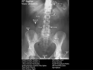
Abdominopelvic Cavity • Abdominal Cavity • Pelvic Cavity P242-fig.4.21
DIVISIONS P242-fig.4.22
Which one of the following is not one of the 9 regions of the abdomen? • Right hypochondriac • Left inguinal or iliac • Epigastric • Right upper • Left lumbar
Which of the following is NOT true concerning the peritoneal cavity? • The peritoneal cavity is a potential space. • The peritoneal cavity contains organs inside of it. • The peritoneal cavity is filled with fluid that lubricates its contents. • The parietal and visceral peritoneum are linings of the peritoneal cavity.
The usual location for an appendectomy incision is the: • left lower quadrant • left upper quadrant • right lower quadrant • right upper quadrant
You were asked to assist in a surgical operation on a young patient to treat an ulcer in the first part of the duodenum. You would expect that the surgeon will approach the ulcer by doing an anterior abdominal wall incision in the following region:
Epigastric • Left inguinal • Left lumbar • Right hypochondrial • Hypogastric
Abdominal wall Anterolateral abdominal wall Posterior abdominal wall
Skin Superficial fascia Deep fascia Muscles Transversalis fascia Extraperitoneal fascia Peritoneum LAYERS
Superficial fascia • Camper’s fascia • Scarpa's fascia P245-fig.4.25~4.26
SUPERFICIAL ARTERIES • Lateral • Posterior intercostal a. • Subcostal a. • Lumbar a. • Median • Epigastric a. • hypogastric a. • Inferior • Superficial epigastric a. • Superficial iliac a. P255-fig.4.39
Superficial veins lateral thoracic subclavian thoracoepigastric portal paraumbilical S epigastric femoral S circumflex iliac
INNERVATIONS • Intercostal Nerve • T7-T12 • 10th Intercostal Nerve
MUSCLES Anterior Group Lateral Group • External Oblique • Internal Oblique • Transversus • Rectus Abdominis • Pyramidalis
RECTUS ABDOMINIS • Tendinous Intersection (3) • Linea Semilunaris
Arteries • 5 intercostal arteries • subcostal arteries • 4 lumbar arteries • Superior epigastric artery—internal thoracic artery • Inferior epigastric artery -external iliac artery • Deep iliac circumflex artery- external iliac artery
Innervations • Intercostal n. • Anterior cutaneous branch • Lateral cutaneous branch
T7-12 thoracic n. • Iliohypogastric n. • Ilioinguinal n. • Genitofemoral n.
Umbilical Folds • Median -- median umbilical lig. • Medial -- chorda arteriae umbilicalis • Lateral -- inferior epigastric a. & v.
INCISIONS • Longitudinal • Midline • Paramedian • Transrectal • Oblique • Subcostal • McBurney’s • Transverse • Pfannenstiel • Combined • Thoracal-abdominal
The inferior border of the rectus sheath posteriorly is called the: • Falx inguinalis • Inguinal ligament • Internal inguinal ring • Arcuate line • Linea alba
Following an emergency appendectomy your patient complained of having paresthesia (numbness) of the skin at the pubic region. The most likely nerve that has been injured during the operation is: • Genitofemoral • Iliohypogastric • Subcostal • Spinal nerve T10 • Spinal nerve T9
An obstetrician decides to do a Caesarean section on a 25-year-old pregnant woman. A transverse suprapubic incision is chosen for that purpose. All of the following abdominal wall layers will be encountered during the incision EXCEPT the:
Anterior rectus sheath • Posterior rectus sheath • Rectus abdominis muscle • Skin and subcutaneous tissue • Transversalis fascia, extraperitoneal fat, and peritoneum
Surgical approaches to the abdomen sometimes necessitate a midline incision between the two rectus sheaths, i.e., through the: • Linea aspera • Arcuate line • Semilunar line • Iliopectineal line • Linea alba
The internal thoracic artery is sometimes surgically cut near the caudal end of the sternum and used to supply blood to a region of the heart. In these cases, maintenance of adequate blood flow to the rectus abdominis may be dependent on increased flow through which artery?
Superficial epigastric • Inferior epigastric • Umbilical • Superficial circumflex iliac • Deep circumflex iliac