Immune responses
Immune responses. Dr Kathy Triantafilou University of Sussex School of Life Sciences. Bacterial infections. Bacteria enter the body through either: a number of natural routes respiratory track gastrointestinal track genitourinary track unnatural routes openings by breaks in the skin

Immune responses
E N D
Presentation Transcript
Immune responses Dr Kathy Triantafilou University of Sussex School of Life Sciences
Bacterial infections • Bacteria enter the body through either: • a number of natural routes • respiratory track • gastrointestinal track • genitourinary track • unnatural routes • openings by breaks in the skin • openings by breaks in mucous membranes
Host defense • Different levels of host defense are enlisted depending on: • the number of organisms • if the inoculum size and virulence are low, then localised tissue phagocytes maybe able to eliminate the bacteria (innate immune system) • virulence of the organisms • Larger inoculums or organisms with greater virulence tend to induce and adaptive, specific immune response
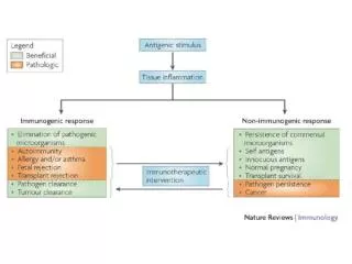
Extracellular bacteria • Extracellular bacteria are pathogenic because: • they induce a localised inflammatory response • they produce toxins • the toxins, endotoxin (LPS) or exotoxin can be cytotoxic • may cause pathogenesis by other ways: • endotoxins (LPS) which are components of bacterial cell wall stimulate can cause oversecretion of cytokines (septic shock) • toxin secreted by diphtheria blocks protein biosynthesis by the cell
Extracellular bacteria • Humoral immune response is the main protective response against extracellular bacteria • Antibodies that bind to antigens on the surface of a bacterium can together with C3b component of complement increase phagocytosis and enhance clearance of the bacterium • Complement activation can lead directly to lysis of the organism • Complement activation can induce production effector molecules that help in developing an inflammatory reponse (complement split products)
Intracellular bacteria • Innate immunity is not effective against intracellular bacterial pathogens • Intracellular bacteria can activate NK cells • NK cells provide an early defense against these bacteria • Intracellular bacteria induce cell-mediated immune response (delayed-type hypersensitivity) • Cytokines are secreted by CD4+ T-cells, notably IFN-g which activates macrophages to kill ingested pathogens
Steps in bacterial infection • Attachment to host cells • Proliferation • Invasion of host tissue • Toxin-induced damage to host cells
Attachment • Bacteria have surface structures that enhance their ability to attach to host cells (pili-long hairlike projections) • Bordetella pertussis secrete adhesion molecules that attach to both the bacterium and the epithelial cells of the upper respiratory track • Secretory IgA antibodies specific for such bacterial structures can block bacterial attachment to mucosal epithelial cells (main host defense against bacterial attachment)
Bacterial Evasion • Some bacteria (e.g. Neisseria gonorrhoea, Haemophilus influenzae, and Neisseria meningitidis) evade the IgA response by secreting proteases that cleave secretory IgA at the hinge region • Some bacteria evade the IgA response of the host by changing their surface antigens (e.g. in N. gonorrhoeae the protein component of pilin has a highly variable structure) • variation in the pilin amino acid sequence is generated by gene rearrangement • this contributes to the pahtogenicity of N. gonorrhoeae by allowing it to bind to epithelial cells
Bacterial Evasion • Some bacteria possess surface structures that serve to inhibit phagocytosis • Streptococcus pneumoniae has a polysaccharide capsule that prevents phagocytosis (there are 84 serotypes that differ in the capsular polysaccharide) • Streptococcus pyogenes has a surface projection called the M protein which inhibits phagocytosis • Some staphyloccoci are able to assemble a protective coat from host proteins. These bacteria secrete a coagulase enzyme that precipitates a fibrin coat around them, shielding them from phagocytic cells
Bacterial evasion • In some gram-negative bacteria long side chains on the lipid A of the LPS help to resist complement-mediated lysis • Pseudomonas secretes an enzyme, elastase, that inactivates both the C3a and C5a anaphylatoxins, thus diminishing localised inflammatory reactions • Some bacteria escape host defense mechanisms by their ability to survive within phagocytic cells
Bacterial evasion • Listeria monocytogenes escapes from the phagolysosome to the cytoplasm, which is a more favorable environment for their growth • Mycobacterium avium blocks lysosomal fusion with the phagolysosome, and some mycobacteria are resistant to the oxidative attack that takes place within the phagolysosome • Salmonella has evolved the ability to enter into cells that are normally nonphagocytic. On contact with the cells Salmonella delivers a number of bacterial effector proteins into the host cell cytosol, interfering with the actin cytoskeleton of the cell and thus gains entry (Galan and Zhou, 2000)
Contribution of the immune system to bacterial pathogenesis • In some cases, disease is not caused by the bacterial pathogens, but by the immune response to the pathogen: • septic shock (oversecretion of cytokines) • food poisoning • toxic-shock syndrome • exotoxins produced by the pathogens function as superantigens which can activate all T-cells leading to overproduction of cytokines
General characteristics • Multiply within living cells by using the biosynthetic machinery of the host • Contain a single type of nucleic acid, either DNA or RNA • Contain a protein coat (the capsid) consisting of individual protein units (capsomeres) • May contain a host derived lipid membrane (the envelope) through which may be inserted viral proteins (spikes) • Small filterable through bacteriological filters
Virus morphology • Helical (e.g. bacteriophage M13) • Polyhedral/Cubic (e.g. poliovirus) • Enveloped (e.g. HIV) • Complex (e.g. poxviruses)
Major virus families Family Envelope Example Adenoviridae No Adenovirus Arenaviridae Yes Lassa fever virus Bunyaviridae Yes Hantaan Calicividae No Norwalk virus Coronaviridae Yes 229E Filoviridae Yes Marburg Flaviviridae Yes Hepatitis C virus Hepadnaviridae No Hepatitis B virus Herpesviridae Yes Cytomegalovirus Orthomyxoviridae Yes Influenza Papovaviridae No Papillomavirus Paramyxoviridae Yes Respiratory syncytial virus Parvoviridae No RA1 Picornaviridae No Coxsackievirus Poxviridae Yes Monkeypox virus
Major virus families Family Envelope Example Reoviridae No Rotavirus Retroviridae Yes HIV Rhabdoviridae Yes Rabies Togaviridae Yes Rubella
Infectious cycle of viruses • Attachment, using cell surface receptors • Cell entry • Nucleic acid and protein synthesis • Assembly of virions • release of virus particles from host cell
Virus Receptors • It has been clear for many years that viruses which propagate within vertebrate hosts have adapted many strategies in order to infect host cells • One of the first steps in a viral infection is the binding of the virus to cell surface molecules.This interaction plays a key role in the multiplication cycle. • Entry of viruses into cells is a complex multi-step process and for several viruses cell attachment and internalisation are distinct steps
Entry of viruses into cells is a complex multi-step process • HIV-1 -CD4, CXCR4, CCR5 • Coxsackie B viruses -CD55, CAR protein, 100kDa nucleolin protein • HSV - Heparan sulphate, PRR1 and PRR2 • CAV-21 CD55, ICAM1 • Adenovirus -CAR protein, avb3, avb5, b2 integrins
Virus evasive strategies • The evolution to use multiple complexes of receptors for their cell attachment and entry provides viruses with cell tropism for different tissues and organs -HIV1 initially binds CD4 while CXCR4 or CCR5 are required for cell entry in T cells or macrophages respectively - Adenovirus binds CAR protein, while uses b2 integrins for entry into blood cells,and avb3 or avb5 integrins for entry in other tissues.
Viral evasion strategies • Infection of sites not accessible to the immune system -Infection of central nervous system (neurons do not express MHC) - Epithelial surfaces with limited T cell access • Antigenic Variation -Viruses undergo mutations at high frequency • Viral escape of T cell recognition -Mutations of the sequences encoding the epitope seen by the TCR • Suppression of MHC molecules -Interference with the presentation of viral peptides by the host
Strategies to induce immunosuppression • Infect T and B cells and abrogate their function (e.g. HBV infects B and T cells, HSV infects T cells, EBV infects B cells) • Destroy antigen presentation cells (e.g. CMV, HIV) • Down regulate viral protein expression (e.g. HSV) • Infect cells lacking MHC class I (e.g. measles virus) • Production of viral proteins that interfere with MHC class I (e.g. CMV, HSV)
Diphtheria (Corynebacterium diptheriae) • Diptheria is an example of bacterial disease caused by a secreted exotoxin (immunity can be induced by immunization with an inactivated toxoid) • It was first described by Klebs in 1883 and was shown a year later by Loeffler to cause diphtheria in guinea pigs and rabbits • Autopsies of the infected animals revealed that the damage from the bacterium was widespread. This led Loeffler to speculate that the manifestations of the disease were caused by a toxic substance secreted by the organism
Diphtheria • His hypothesis was validated in 1888, when Roux and Yersin produced the disease in animals by injecting a sterile filtrate from a culture of the bacterium • In 1923, Ramon found that by exposing the toxin to heat and formalin rendered it nontoxic but did not destroy its antigenicity • In 1989, the CDC in the States only reported three cases in the whole of the U.S.
Diphtheria • The disease is spread from one individual to another by airborne respiratory droplets • The bacteria colonises the nasopharyngeal tract, remaining in the superficial layers of the mucosa • Growth of the bacterium causes little tissue damage • The virulence of the organism is due completely to its potent exotoxin • The toxin causes destruction of the underlying tissue, resulting in the formation of a tough fibrinous membrane (pseudomembrane)
Diphtheria • The pseudomembrane is composed of fibrin, white blood cells, and dead respiratory epithelial cells • The membrane itself can cause suffocation • The exotoxin also is responsible for widespread systemic manifestations (pronounces myocardial damage and neurologic damage) • The toxoid is administered together with tetanus toxoid and inactivated Bordetella pertussis in a combined vaccine that is given to children of 6-8 weeks
Tuberculosis (Mycobacterium tuberculosis) • Tuberculosis is the leading cause of death in the world from a single infectious agent (killing about 3 million people every year) • About 1.79 billion people (1/3 of the world’s population) are infected with M. tuberculosis • Re-emerged in the 1990’s particularly in the cities where HIV-infection levels are high • Infection usually results from inhalation of small droplets of respiratory secretions containing a few bacilli
Tuberculosis • The inhaled bacilli are ingested by macrophages and are able to survive and multiply intracellularly by inhibiting formation of phagolysosomes, when the infected macrophaes lyse, large numbers of bacilli are released • A cell-mediated CD4+ T-cell response is responsible for much of the tissue damage in the disease • CD4+ T-cell activity is the basis for the tuberculin skin test to the purified protein derivative (PPD) from M. tuberculosis
Tuberculosis • In pulmonary infection, CD4+ T-cells are activated within 2-6 weeks after infection inducing the infiltration of activated macrophages • These cells “wall off” the bacteria inside a granulomatous lesion called the tubercle • A tubercle consists of lymphocytes and a collection of activated macrophages • The massive activation of macrophages that occurs within tubercles often results in the concentrated release of lytic enzymes
Tuberculosis • These enzymes destroy nearby healthy cells, resulting in circular regions of necrotic tissue, which eventually form a lesion with a caseous (cheese-like) constistency • As these lesions heal, they become calcified and are readily visible by X-rays, where they are called Ghon complexes • The activated macrophages suppress proliferation of the phagocytosed bacilli and thus the infection is contained
Therapy • Several drugs (sometimes used in combination), isoniazid, rifampin, streptomycin, pyrazinamide and ethambutol • The intracellular growth of M. tuberculosis makes it difficult for drugs to reach the bacilli • Drug therapy must be continued for at least 9 months to eradicate the bacteria • The vaccine for M. tuberculosis is the attenuated strain of M. bovis called BCG (Bacillus Calmetter-Guerin)
Lyme Disease (Borrelia burgdorferi) • In 1975, about 60 cases of a newly observed disease were reported in Lyme, Connecticut • The disease symptoms included unexplained “bull’s eye” rashes, headaches, and arthritis • In some cases, severe neurologic complications developed: excruciating headaches, meningitis, loss of memory, and mood swings • No causative agent was isolated until 1977, Willy Burgdofer found that the patients were bitten by ticks
Lyme Disease • It was found that the tick was teeming with a new species of gram-negative spirochete, which was subsequently named Borrelia burgdorferi • Since the tick takes a blood meal, B. burgdoferi enters the bloodstream • Lyme disease begins with a characteristic rash, appears as a bull’s eye 10-50 cm in diameter • After the rash, arthritic and neurologic symptoms develop • The disease can be successfully treated with broad-spectrum antibiotics such as penicillin
Lyme disease • Antibodies to a protein associated with the flagella of B. burgdorferi can often be detected after infection • These antibodies do not confer protection against infection, but it contributes to the pathogenesis of Lyme disease • Immune complexes (antigen-antibody) are thought to result in a type III hypersensitivity reaction that causes the arthritic symptoms (complexes are deposited at the joints)
Meningitis • Caused by Neisseria meningitidis, Haemophilus influenzae, and Streptococci • Usually bacteria colonize the throat (sore throat), where they gain access into the bloodstream (septicemia). After replication in the bloodstream, they reach the meninges (lining of the brain) • Therapy: vaccine against Haemophilus influenzae (very effective)
Vaccines • Neisseria has five main Groups - A,B,C, W135 and Y • Most UK meningococcal disease is caused by groups B and C • There are combined vaccines for group A and C, that can give some protection • Effective vaccines for Group B are still some years away (which accounts for 65-70% of the cases)
Autoimmunity • Inappropriate response of the immune system against self-components • First observed by Paul Ehrlich early in this century, and he termed the condition “horror autotoxicus” • Not all self-reactive lymphocytes are deleted during T and B-cell development • Self-reactive lymphocytes are re-circulating, their activity regulated by clonal anergy or clonal suppression
Autoimmunity • The damage to self-cells or organs is caused by: • antibodies • Autoimmune hemolytic disease (antigens on red blood cells are recognised by auto-antibodies) • Hashimoto’s thyroiditis (antibodies attack thyroid peroxidase or thyroglobulin and cause severe tissue destruction • T-cells • rheumatoid arthritis • insulin-dependent diabetes mellitus
Hashimoto’s thyroiditis • An individual produces auto-antibodies and sensitised TDTH cells specific for thyroid antigens • The DTH response is characterised by an intense infiltration of the thyroid gland by lymphocytes, macrophages and plasma cells which form germinal centers • Antibodies are formed to a number of thyroid proteins, including thyroglobulin and thyroid peroxidase • Binding of these antibodies to thyroid tissue interferes with the iodine uptake and leads to decreased production of thyroid hormones
Autoimmune anemias • Include pernicious anemia, autoimmune hymolytic anemia and drug-induced hemolytic anemia • Pernicious anemia is caused by auto-antibodies to a membrane bound intestinal protein on gastric cells (intrinsic factor), that facilitates the uptake of vitamin B12 from the small intestine • In the absence of B12, which is necessary for hematopoiesis, the number of functional mature red blood cells decreases below normal • It is treated by injections with B12
Autoimmune anemias • An individual with autoimmune hemolytic anemia makes auto-antibodies to RBC antigens • This triggers complement-mediated lysis or antibody-mediated opsonization and phagocytosis of RBCs • One form of autoimmune anemia is drug-induced: certain drugs (such as penicillin) interact with RBCs and the cells become antigenic
Goodpasture’s syndrome • Auto-antibodies specific for certain basement-membrane antigens bind to the basement of the membranes of the kidney and the alveoli of the lungs • This leads to complement activation and direct cellular damage as well as an inflammatory response mediated by the build-up of complement split products • Tissue damage leads to kidney damage and pulmonary hemorrhage • Death ensues often within several months
Insulin-dependent Diabetes Mellitus (IDDM) • Autoimmune attack of the pancreas • The attack is directed against specialised insulin-producing cells (beta cells) that are located in spherical clusters called the islets of Langerhans • The autoimmune attack destroys the beta cells, resulting in decreased production of insulin and consequently increased levels of blood glucose