Download
1 / 48
480 likes | 589 Vues
Normal Immune Responses, Hypersensitivities, and Allograft Rejection. Normal immune response. Protection against infectious disease Innate immunity epithelial barriers phagocytic neutrophils and macrophages natural killer (NK) cells circulating plasma proteins (complement, clotting)

E N D
Normal Immune Responses, Hypersensitivities, and Allograft Rejection
Normal immune response • Protection against infectious disease • Innate immunity • epithelial barriers • phagocytic neutrophils and macrophages • natural killer (NK) cells • circulating plasma proteins (complement, clotting) • Adaptive immunity— “the immune response” • cellular • B-cells and plasma cells • helper (Th) and cytotoxic (CTL) T-cells • humoral • antibodies • cytokines
Lymphocytes and Receptors • Tcells—TCRs recognize MHC-Ag complexes • Class I MHC universally expressed • HLA-A, HLA-B, HLA-C single a-b2 microglobulin dimer • Ag is peptide processed in cytoplasm by proteasome from intracellular microbe (B,V) or tumor-associated protein • TCR a b dimer complexed with z, CD3 and CD8, CD28 • Class II MHC restricted to DC, MF, B-cells • HLA-DP, HLA-DQ, HLA-DR a–b dimer • Ag is lysozome-processed peptide from extracellular microbe (B,E) protein • TCR a b dimer complexed with z, CD3 and CD4, CD28 • Bcells—IgM complexes with Iga, Igb and CD40 or CD21 coreceptor • NK cells—class I MHC inhibitory receptors with CD16 (IgG Fc receptor) and NKG2D coreceptors
Antigen-Presenting Cells • Dendritic Cells • Dendritic Cells (DCs) • located in T-cell zones of lymphoid tissues to present antigens to circulating T cells • express high levels of class II MHC and T-cell costimulatory molecules • resident in tissues, e.g. Langerhans cell of the epidermis and in the interstitium of many nonlymphoid organs, e.g. heart and lungs • Follicular Dendritic Cells (FDCs) • located in the germinal centers of lymphoid follicles in the spleen and lymph nodes to display antigens to activated B cells • express IgG Fc and complement receptors to efficiently trap antigen bound to antibodies and complement • Macrophages • phagocytosed microbes and protein antigens presented as peptide fragments to T cells • B cells • present peptides to Th cells and receive signals that stimulate antibody responses to protein antigens
Pattern recognition receptors • DCs and macrophages respond to pathogen-associated and damage-associated molecular motifs • Toll-Like Receptors (10 in human) • membrane receptors on outer plasma membrane and vessicle membranes • TLR2 for Gram positives, TLR3 for fungi, TLR4 for Gram negatives, TLR9 for viral and bacterial DNA (CpG) • NLRs (NOD-like receptors) (at least 20) • Cytosolic proteins bind a variety of microbial products • NLRC-type have caspase activation domain • NLRP-type have pyrin domain • Activate caspases and NF-kB • Affect gene expression via NF-kB and MAP kinase cascades • Release IL-1 and other proinflammatory cytokines
Tissues of the Immune System • Generative Lymphoid Organs • thymus, where T cells develop • bone marrow, where all blood cells are produced and where B cells mature • Peripheral Lymphoid Organs • lymph nodes, where lymph-borne antigens are trapped by FDCs and where DCs concentrate • spleen, where blood-borne antigens are trapped by FDCs and where DCs macrophages concentrate • T lymphocytes and B lymphocytes are segregated into different regions
MHC is HLA HLA and Disease Association
Hypersensitivity Reactions • Exposure to antigen results in sensitivity • Repeat exposure may result in pathologic hypersensitivity • Both exogenous and endogenous antigens may elicit hypersensitivity • Hypersensitivity is an imbalance between effector and control mechanisms of immune responses • Development of hypersensitivity is often associated with the inheritance of particular susceptibility gene
Types of hypersensitivity reactions • Type I — immediate • immunologic reaction occurs within minutes of antigen binding to antibody bound to mast cells in individuals with prior sensitization • Type II — Ab reaction to bound Ag • caused by antibodies that react with antigens present on cell surfaces or in the extracellular matrix • Type III — Ab complex with circulating Ag • antigen-antibody complexes deposited on vessel walls cause inflammation and tissue damage • Type IV — delayed-type • initiated by antigen-activated (sensitized) T cells
Immediate hypersensitivity • Presentation of antigen to naive Th cells • Naive cells differentiate into Th2 cells • Th2 cells produce cytokines upon subsequent encounter with the antigen • IL-4 stimulates B cell class switching to IgE and promotes additional Th2 cell development • IL-5 promotes development and activation of eosinophils • IL-13 enhances IgE production and stimulates mucus secretion by epithelial cells • Mast cells and basophils bind IgE
IgE cross-linking activates mast cells • Hypersensitivity mediated by IgE-dependent activation of mast cells upon re-expsure to antigen • mast cells are bone marrow–derived • abundant near blood vessels, nerves and subepithelial tissues • cytoplasmic membrane-bound granules contain active mediators and acidic proteoglycans that bind basic dyes • activated by cross-linking of high-affinity IgE Fc receptors • triggered by complement C5a and C3a • Basophils also have cell surface IgE Fc receptors and cytoplasmic granules • circulate in the blood in extremely small numbers but can be recruited to inflammatory sites
Mast cell degranulation • Preformed mediators released from granules • Vasoactive amines • histamine causes intense smooth muscle contraction, increased vascular permeability, and increased mucus secretion by nasal, bronchial, and gastric glands • Enzymes • neutral proteases (chymase, tryptase) and several acid hydrolases • Proteoglycans • Heparin (anticoagulant) and chondroitin sulfate package and store the amines in the granules
Mast cell lipid mediators • Synthesized after activation of PLA2 releases AA from plasma membrane • Leukotrienes • LTC4 and LTD4 - several thousand times more active than histamine in increasing vascular permeability and causing bronchial smooth muscle contraction • LTB4 is highly chemotactic for neutrophils, eosinophils, and monocytes • Prostaglandin D2 • causes intense bronchospasm as well as increased mucus secretion • Platelet-activating factor (PAF) • causes platelet aggregation, release of histamine, bronchospasm, increased vascular permeability, vasodilation and is chemotactic for neutrophils and eosinophils • PLA2 dependent, not AA product
Mast cell cytokines • TNF, IL-1, and chemokines (eotaxin, CXCL8) • attract neutrophils, eosinophils, basophils, monocytes • IL-4 • amplifies the Th2 response • IL-3, IL-5, and GM-CSF • support survival of eosinophils • Cell recruitment and survival supports the late-phase response
Systemic Anaphylaxis • Subsequent exposure to minute amounts of antigens in previously sensitized individuals • Hospital acquired • administration of foreign proteins (e.g. antisera), hormones, enzymes, polysaccharides, or drugs (e.g. penicillin) • Community acquired • food allergens (e.g. peanuts, shellfish) or insect toxins (e.g. bee venom) • Within minutes after exposure: • itching, hives, skin erythema, contraction of respiratory bronchioles and respiratory distress • Followed shortly by: • vomiting, abdominal cramps, diarrhea, and laryngeal obstruction due to edema • Within an hour • circulatory shock due to massive edema and hypovolemia
Atopy • Chronic, localized, anaphylactic-like responses to antigens • Asthma • Eczema (dermatitis) • Allergic rhinitis • Urticaria (hives and wheals) • Mediated by mast cells and eosinophils with similar symptoms • bronchoconstriction, inflammation, itching, edema
Type II hypersensitivity • Opsonization and Phagocytosis • Mechanism involves IgG or IgM • Cells opsonized by IgG antibodies are recognized by phagocyte Fc receptors • Opsonization activates the complement system by the classical pathway • Complement activation forms the membrane attack complex • Osmotic lysis of cells • Antibody-dependent cellular cytotoxicity (ADCC) • cell lysis proceeds without phagocytosis • NK cells
Destruction of blood cells • Transfusion reactions • cells from incompatible donor are opsonized by preformed antibody in the host • Erythroblastosis fetalis • hemolytic disease of the newborn • mother-fetus antigenic difference (e.g. Rh factor) • maternal IgG crosses the placenta to cause destruction of fetal red cells • Autoimmune hemolytic anemia, agranulocytosis, and thrombocytopenia • individuals produce antibodies to their own blood cells • Drug reactions • drug binds to RBC, acts as hapten for Ig activation
Type III Hypersensitivity • Formation of immune complexes • abundant, novel protein antigen triggers immune response resulting in antibodies • secreted antibodies react with the antigen still present in the circulation forming antigen-antibody complexes • Deposition of immune complexes • organs where blood is filtered at high pressure to form other fluids, like urine and synovial fluid, are most affected • Tissue injury • acute inflammatory reaction with complement-fixing antibodies (i.e., IgG and IgM) leukocyte Fc receptor-antibody complexes induce pathologic lesions
Immune complex injury • Principal morphologic manifestations • acute necrotizing vasculitis • necrosis of the vessel wall • immune complexes, complement, and plasma protein produce eosinophilic, fibrinoid necrosis • Chronic serum sickness • repeated exposures to antigen • SLE persistent antibody responses to autoantigens • Local Immune Complex Disease • Arthus reaction • localized area of tissue necrosis resulting from acute immune complex vasculitis in the skin
Type IV hypersensitivity • Initiated by antigen-activated (sensitized) T cells • Delayed-type hypersensitivity (DTH) • CD4+ Th1 cell cytokines recruit macrophages • induced by environmental and self-antigens • Direct cell cytotoxicity • CD8+ CTLs cause tissue damage • frequently follow viral infections • Many autoimmune diseases are type IV hypersensitivities
Tuberculin reaction • Classic example of DTH • intracutaneous injection of purified a protein-containing antigen of the tubercle bacillus (tuberculin) • reddening and induration of the site appear in 8 to 12 hours, reach a peak in 24 to 72 hours • characterized by the accumulation of CD4+ T cells and macrophages around venules, producing perivascular “cuffing”
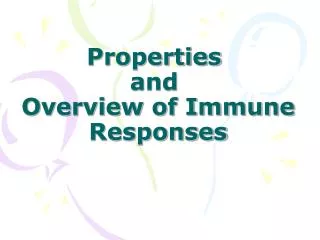
Tb skin test Within 48 to 72 hours, a positive TB skin test is marked by an area of reddish induration greater than 10 mm. It is the induration (firm bump) that is gently palpated that determines the size, not the area of redness. This reaction is slightly larger than the average positive test 17 mm in size. The positive reaction shown here was obtained with a TB skin test performed 20 years after the initial infection.
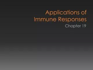
Type 1 DM An islet of Langerhans demonstrates insulitis with lymphocytic infiltrates in a patient developing type I diabetes mellitus. This lesion precedes clinical onset of diabetes mellitus and is rarely observed.
Mechanism of CD4+ T cell DTH • Naive CD4+ T cells recognize peptides displayed by dendritic cells • Differentiation of antigen-stimulated T cells to Th1 or Th17 cells is driven by the cytokines produced by APCs • IL-12 induces differentiation of CD4+ T cells to the Th1 subset which makes IFN-γ • IL-1, IL-6 and IL-23 with TGF-β stimulate differentiation of T cells to the Th17 subset • Previously activated T cells respond to subsequent exposure • Th1 cells secrete cytokines, mainly IFN-γ • IFN-γ–activated macrophages express more class II MHC, secrete TNF, IL-1, and chemokines, and produce more IL-12 • Activated Th17 cells secrete IL-17, IL-22, chemokines, and other cytokines that recruit neutrophils and monocytes to the reaction • Macrophages promote fibrosis and damage
Mechanism of CD8+ cytotoxicity • Naïve CD8+ recognize Ag in the context of MHC-class I on cell surfaces • Differentiated CTLs contain perforin-granzyme protease complexes that induce apoptosis in target cells • This mechanism plays a role in T1DM and transplant rejection