Metastasis
Metastasis . How the breakdowns of normal cell adhesions and stasis make cancers much more dangerous. Survival data show that prognosis gets worse when distant metastasis has occurred. . Metastasis is important for cancer medicine.

Metastasis
E N D
Presentation Transcript
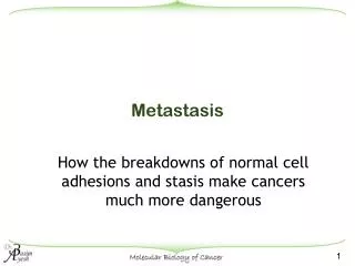
Metastasis How the breakdowns of normal cell adhesions and stasis make cancers much more dangerous
Survival data show that prognosis gets worse when distant metastasis has occurred. Metastasis is important for cancer medicine
Fraction of all cancer patients surviving months after diagnosis, based on the presence (+) or absence (-) of distant metastasis
Therefore we need to understand metastasis • Cell adhesion is a complex process that depends on many different molecules • Destruction of the cell adhesion machinery can result from “loss of function” mutations in one or more of the relevant adhesion molecules
Both cell-cell interactions and cell-matrix interactions play an important role during the invasive cascade. • Connections through cell-adhesion molecules, integrins, and cadherins, stabilize tissue integrity • Loss or alteration of these cell surface proteins is associated with increased metastatic potential.
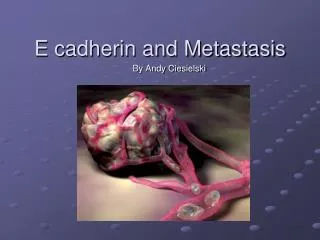
Cadherins • Cadherins are transmembrane glycoproteins that mediate extracellular calcium-dependent cell-cell interactions • E-(epithelial) cadherin, the most extensively studied, is involved in epithelial cell-cell communication • Bind the cytoskeleton of one cell to that of its neighbors, forming a unit. • This coupling contributes to the mechanical integrity of a tissue.
Localization of Cadherin revealed by fusing thisprotein with the Green Fluorescent Protein (GFP)
Cells also bind to one another by cell adhesion molecules that do not bind Ca. • Here are 4 CAMs intercellular (cell adhesion molecule), showing their extracellular folds, membrane spanning domains, and ability to dimerize, and form cell-cell bonds.
Desmosomes • Make mechanical links between the intermediate filaments of one cell and its neighbors. • This contributes significantly to the mechanical strength of epithelia.
Integrins • Integrins are a family of transmembrane glycoproteins that are expressed by the cell as α/β heterodimers • Membrane proteins that bind to the Extracellular Matrix (ECM). • The integrins make bonds between the actin cytoskeleton and the fibers of the ECM, such as collagen and fibronectin.
Originally identified as cell adhesion molecules, • Recognized as signaling molecules for regulation of apoptosis, gene expression, cell proliferation, invasion and metastasis, and angiogenesis
What happens when cell junctions are broken or weakened? • Watching the direct result of mutations that induce metastasis is not easy to do, because you don’t know when the mutation will occur • Thus, we can either compare properties of metastatic cells with those of normal cells or set up experiments that knock out known components of cell adhesion and see how their loss compares with the onset of metastasis
The junctions between normal cells are strong, but loss of normal adhesion molecules can reduce the strength of cell-cell interactions. • In (a-c) significant force is required to pull normal cells apart. • In (d-f) metastatic cells are seen to require much less force to pull them apart.
Cell junctions can fail as a result of mutations that alter CAMs • A loss-of-function allele of a Cadherin, a Desmocollin, or a Ca++-independent CAM can lead a cell to a lose its adhesion to proper neighbors and start to wander • The wandering cell must then get from the local stroma to more distant sites for true metastasis growth
The most common LOF mutations for carcinomas are in Cadherins • In metastatic prostatic carcinomas, cadherins are down-regulated, as measured by PCR • This down-regulation is often a result of a single nucleotide mutation in the DNA 5’ to the structural gene, presumably its promotor • Snail is a transcription factor that down-regulates cadherin expression. Its hyper-activity can lead to lowered levels of CAMs
Loss of adhesion is necessary, but not sufficient for metastatic behavior • To get to a site of metastatic growth, a cells must also wander from its initial location • At first site, cell wandering would appear to be a novel property of cancer cells. • There are many circumstances in which cell migration is a normal and even essential part of cell behavior
When cell junctions are broken by a wounding process, cell migration is induced as a normal part of wound healing • In vivo, our ability to close a cut depends on cell migration and proliferation • These processes can be modeled in cell culture • Confluent layers of epithelial and fibroblastic cells are comparatively static • With wounding, cells become motile and division will often ensue.
Breaking important cell-cell junctions can occur by LOF mutations, but it takes several losses to let a cell loose • The probability of acquiring one mutation in a normal mammalian cell is about 1 in 100 million cell divisions • For a mutation to become homozygous, additional improbable events must occur • Look, therefore, for simple ways to bring about complex events, like loss of cell adhesion • The easiest is to secrete proteases that cut the proteins that normally hold cells together.
Cadherin function can be down-regulated by post-translational effects • Calpain, an intracellular protease, can cut cadherin down to a 100 kD polypeptide that lacks its binding sites for beta-catinin • Extracellular proteases, like metalo-protease can cut cadherin and related CAMs, reducing their effectiveness in holding tissues together • All such effects promote metastasis
Mutations that lead to the secretion of proteases are particularly dangerous • Collagenases will loosen and degrade some ECM • There are several metaloproteases that are important for normal tissue morphogenesis and remodeling; examples: stromelysins 1 and 3. • Over expression and secretion of such enzymes can degrade cadherins and other cell adhesion molecules, and can modify basal lamellas making it easier for circulating cells to pass through • Overexpression of Stromelysin 1 is sufficient to make a cell metastatic. Thus, one GOF mutation can turn a hypertrophic cell into a metastatic cell
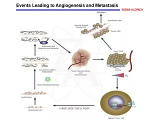
When cell junctions fail, cells can wander, but to be dangerously metastatic they do not wander without logic • First job: cross the BL. For this, proteases (particularly collagenases) are essential • Once the cell has gotten into the stroma, (connective tissue below the BL) there are pathways that can facilitate cell migration • Blood vessels are one such conduit • Lymph vessels are another
The lymph vessels, or “lymphatics” provide a network of pathways for metastatic movements
Epithelial cells that can cross the BL and migrate through connective tissue can also enter blood vessels and follow a fast-track to distant locations
Travel in blood is, however, dangerous for the metastatic cell • Fast flow of blood implies sheer, which can destroy the metastatic cells. • Indeed, correlations between tumor cells in blood and frequency of metastatic growth are not good. • This emphasizes the difficulties faced by tumor cells in accomplishing “extravasation” • Formation of clots (involving platelet activation) can be important for extravasation, but endothelial cells have receptors for cell surface molecules that can bind a metastatic cell in place, giving it time to pass out of the capillary, just as in normal inflammation
Best evidence for blood as a pathway for metastasis is some of the common patterns of metastasis • Tumors in the lung commonly metastasize to the brain. Blood flows from the lung to the heart (return of “pulmonary circulation loop”, then a significant amount goes to the brain. • Intestinal tumors frequently metastasize to the liver (remember the hepatic portal system) • Thus, blood is likely to play an important part in the “logic of metastasis”
Once the tumor cells has escaped from the vasculature, a site of metastatic growth can be established • Affinity of the metastatic cell for local ECM may provide another chemical logic that helps to define sites of frequent metastasis • CAMs that grant cell-cell adhesion specificity can also help to define where metastatic growth will occur • The hospitality of an environment for new growth (degree of vascularization, etc.) can also make a difference