Nephrolithiasis
Nephrolithiasis. Cynthia Denu-Ciocca, M.D. Epidemiology. Incidence 1:1000 per year Peak onset 20 - 35 years of age Male:Female 3 - 4 : 1. Epidemiology.

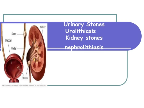
Nephrolithiasis
E N D
Presentation Transcript
Nephrolithiasis Cynthia Denu-Ciocca, M.D.
Epidemiology • Incidence 1:1000 per year • Peak onset 20 - 35 years of age • Male:Female 3 - 4 : 1
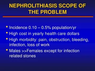
Epidemiology • In their lifetime 2 - 5% of the Asian population 8 - 15% of North Americans and Europeans 20% of Saudi Arabians will develop a kidney stone
Epidemiology • Soucie et al performed a cross sectional study to investigate the geographic variability in the rates of stone formation • 1,167,009 men and women in the U.S. • Stones were 2x as prevalent in the southeast • Ambient temps and sunlight indices were independent predictors of stone formation
Epidemiology • Curhan et al studied the influence of FH on stone formation • 17.2% of men vs. 6.4% + family history • 1986 - 1994 : 795 incident cases of stones • RR of stone formation in men with +FH 2.57 (95% CI 2.19 - 3.02)
Epidemiology • Serio and Fraioli confirmed hereditary predisposition • 22.5% of patients who developed stones in Italy between 1993 - 1994 had a positive FH in one or both of their parents
Natural HistoryRecurrence Rates • 40 % in 2 - 3 years • 55% in 5 - 7 years • 75% in 7 - 10 years • 100% in 15 - 20 years
Stone Formation Urine saturation
Stone Formation Urine saturation Supersaturation
Stone Formation Urine saturation Supersaturation Crystal nucleation
Stone Formation Urine saturation Supersaturation Crystal nucleation Aggregation
Stone Formation Urine saturation Supersaturation Crystal nucleation Aggregation Retention and growth
Saturation • Saturation level - specific concentration of a salt = solubility product and no more salt can be dissolved • Crystals form in supersaturated urine • Saturation is dependent on chemical free ion activities of the components of a stone
Chemical free ion activities • concentration of the relevant ions • urine pH • complexation with substances in the urine
Nucleation • Homogeneous - supersaturation reaches a formation product and nuclei form in free solution • Heterogeneous - crystal growth occurs on the surface of dissimilar but complimentary crystal or foreign substances
Clinical Presentation • Renal colic begins suddenly and intensifies over 15 - 30 minutes • Associated with nausea & vomiting • Pain passes from the flank anteriorly to the groin • At the ureterovesicular junction, urinary frequency and dysuria may occur • Microscopic hematuria> 75%, gross - 18%
Patient Evaluation: BasicSingle Stone Former • Stone history • Medical disease: skeletal disease, IBD, UTI’s, HIV, granulomatous disease • Meds: Lasix, glucocorticoids, theophylline, calcium, vitamins A, C, and D
Patient Evaluation: BasicSingle Stone Former • Family history • Lifestyle/occupation • Diet/fluids: protein, coffee, tea, dairy
Patient Evaluation: BasicSingle Stone Former • Physical Examination • Laboratory data • Urinalysis, urine for cystine • Urine culture - • Blood tests • electrolytes, creatinine, calcium, phosphorous, uric acid, intact PTH if calcium elevated
Patient Evaluation: BasicSingle Stone Former • Radiology • KUB • IVP: anatomic abnormality, medullary sponge kidney • Ultrasound • Unenhanced CT
Patient Evaluation: Complete • All patients with “metabolically active” stones, children, people in demographic groups that don’t usually form stones • Metabolically active stone: grow in size, number, or are passed within 1 year of f/up • Basic evaluation and 24 hour urine for volume, calcium, oxalate, sodium, phosphorous, uric acid, citrate, cystine
Patient Evaluation: Complete • Optimal values of 24-hr urine constituents: • volume > 2 - 2.5 L/day • calcium < 4 mg/kg or < 300 mg ,< 250 mg • uric acid < 800 mg , < 750 mg • citrate > 320 mg • sodium < 200 meq • phosphorous < 1100 mg • pH > 5.5 and < 7.0
Calcium oxalate & calcium phosphate Calcium oxalate Calcium phosphate Uric Acid Struvite Cystine 37% 26% 7% 5 - 10 % 10 - 20% 2% Etiology
Calcium Stones • 70 - 75% of all stones • Calcium oxalate - brown, gray, or tan • Calcium oxalate monohydrate - dumbbell • Calcium oxalate dihydrate - pyramidal • Calcium phosphate - white or beige • Calcium phosphate - elongated (brushite)
Hypercalciuria Hypocitraturia Hyperoxaluria Unclassified 40 - 50% 20 - 30% 5% 25% Calcium StonesPathophysiology
Calcium StonesHypercalciuria - Absorptive Calcium Absorption Serum calcium PTH Urinary Calcium
Calcium StonesHypercalciuria - Absorptive • Urine calcium exceeds 250 mg/day in females, 300 mg/day in males • Most common cause of hypercalciuria • familial, 50% of 1st degree relatives, M=F • Patients have a higher incidence of reduced bone mineral density • Caoxalate & Ca++phosphate stones
Calcium StonesHypercalciuria - Absorptive • Pak et al have proposed two subtypes • Type I - hypercalciuria on random and low calcium diets • Type II - normocalciuria on restricted diets • Etiology unknown • Some patients have high serum calcitriol
Rx: Calcium StonesIdiopathic Hypercalciuria • Maintain urine volume > 2 liters/day • Thiazide diuretics • Potassium citrate • Sodium restriction (2 - 3 grams/day) • Protein restriction ( 0.8 - 1.0 g/kg/day)
HypercalciuriaDent’s Disease • Rare, X-linked inheritance • Tubular defect which causes renal phosphate wasting(PO4 2.9 mg/dl) • Stimulates vitamin D, intestinal calcium absorption, urinary calcium • Rx: Phosphorus replacement
Calcium StonesRenal leak Urinary calcium Serum calcium PTH 1,25 (OH)2 D Bone resorption Calcium absorption
Calcium StonesHypercalciuria - Resorptive PTH 1,25 (OH)2 D Bone Resorption Calcium Absorption Serum Calcium Urinary Calcium
Calcium StonesHyperparathyroidism • 85% adenoma, 15% multigland hyperplasia • Urine pH tends to be higher than in idiopathic hypercalciuria so the fraction of calcium phosphate stones is higher • Rx: Parathyroidectomy
Calcium StonesHypocitraturia • Occurs alone (10%) or with other abnormalities (50%) • More common in females • May be idiopathic or secondary • Acidosis reduces urinary citrate by tubular reabsorption
Calcium StonesHypocitraturia • Associated with distal RTA, metabolic acidosis of diarrhea, & consumption of a diet rich in meat • Tend to form calcium oxalate stones (except Type I RTA) • Rx: dietary protein, alkali (K citrate or K bicarbonate), avoid sodium bicarbonate
Calcium StonesRenal Tubular Acidosis • 2/3rds of patients with Type I RTA have nephrocalcinosis or nephrolithiasis or both • Mechanism for stone formation • Hypocitraturia due to acidosis • Hypercalciuria: acidosis bone resorption • Alkaline urine pH: defect in H excretion
Calcium StonesRenal Tubular Acidosis • Form calcium phosphorus stones largely due to the alkaline urine pH which decreases the solubility of calcium phosphate complexes • Rx: Alkali (calciuria, citraturia), thiazides if hypercalciuria persists
Calcium StonesHyperoxaluria • Most patients with calcium oxalate stones excrete normal urinary oxalate: <40 mg/day • Excessive urinary oxalate • Dietary hyperoxaluria • Endogenous hyperoxaluria • Enteric hyperoxaluria
Calcium StonesDietary Hyperoxaluria • Normal people absorb < 5% of dietary oxalate • Foods rich in oxalate (spinach, chocolate, beets, peanuts) can absorption 25 - 50% • Low calcium diets urinary oxalate excretion
Calcium StonesEndogenous hyperoxaluria • Primary hyperoxaluria - enzymatic deficiencies that result in massive oxaluria • Widespread calcium oxalate deposition in tissues ( bone marrow, blood vessels, heart, and renal parenchyma) • Rx: P.O. fluids, pyridoxine and orthophosphate urinary crystallization
A A B B S S O O R R P P T T I I O O N N Calcium StonesEnteric Hyperoxaluria Calcium Fatty Acids Oxalic Acid Calcium Soaps Calcium Oxalates NORMAL
Calcium StonesEnteric Hyperoxaluria A A B B S S O O R R P P T T I I O O N N Calcium Fatty Acids Oxalic Acid Calcium Soaps Calcium Oxalates ENTERIC HYPEROXALURIA
Calcium StonesEnteric Hyperoxaluria • Treatment • Decrease dietary oxalate and fat • Oral Calcium supplements • Cholestyramine • Increase fluid intake • Oral citrate
Calcium StonesHyperuricosuria • Increased frequency of hyperuricosuria (> 800 mg/day , > 750 mg/day) in patients who form calcium stones • Urate crystals serve as a nidus for calcium oxalate nucleation & comprise 4 - 8% of the cores of calcium stones • Rx: Allopurinol, Potassium citrate
Uric Acid Stones • Smooth, white or yellow-orange • Radiolucent • Crystals form various shapes: rhomboidal, needle like, rosettes, amorphous
Uric Acid Stones • Main determinants of uric acid stone formation • Urinary pH < 5.5, urine volume Hyperuricosuria • Genetic overproduction • Myeloproliferative disorders • High purine diet • Drugs