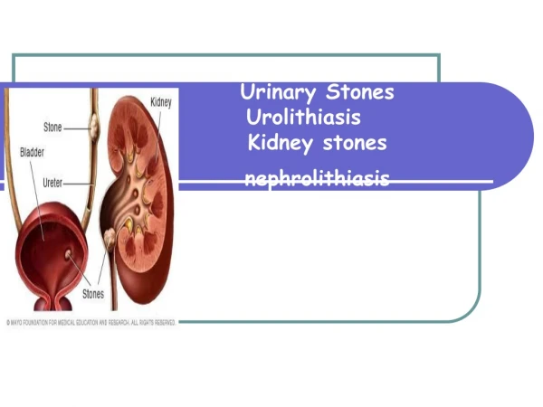
Nephrolithiasis
Nephrolithiasis. Adnan Alsaka M.D. Nephrology Fellow. Nephrolithiasis is estimated to produce medical costs of $2.1 billion per year in the US Incidence is 0.5% Prevalence is 5.2%. More common in Asians and whites than in Native Americans, Africans, African Americans

Nephrolithiasis
E N D
Presentation Transcript
Nephrolithiasis Adnan Alsaka M.D. Nephrology Fellow
Nephrolithiasis is estimated to produce medical costs of $2.1 billion per year in the US • Incidence is 0.5% • Prevalence is 5.2%
More common in Asians and whites than in Native Americans, Africans, African Americans • male-to-female ratio of 3:1 • Stones due to infection (struvite) are more common in women
Most urinary calculi develop in persons aged 20-49 years • An initial stone attack after age 50 years is relatively uncommon
1- supersaturation of the urine by stone-forming constituents Crystals or foreign bodies can act as nidi, upon which ions from the supersaturated urine form microscopic crystalline structures
Randall plaque • is deposition of stone material on a renal papillary calcium phosphate nidus • Calcium phosphate precipitates in the basement membrane of the thin loops of Henle, erodes into the interstitium, and then accumulates in the subepithelial space of the renal papilla • The subepithelial deposits, eventually erode through the papillary urothelium
Hypercalciuria • most common metabolic abnormality • can be subdivided into absorptive, resorptive, and renal-leak categories
Absorptive Hypercalciuria • related to increased intestinal absorption of calcium (associated with excess dietary calcium and/or overactive calcium absorption mechanisms) • the treatment may include modest dietary calcium restriction, thiazide diuretics, oral calcium binders
Resorptive hypercalciuria • related to excess resorption of calcium from bone (i.e., hyperparathyroidism) • Treatment requires parathyroidectomy
Renal-leak hypercalciuria • less common than absorptive hypercalciuria • related to an inability of the renal tubules to properly reclaim calcium in the glomerular filtrate • Usually associated with secondary hyperparathyroidism and is best managed with thiazide diuretics
Indiscriminate dietary calcium restriction is not advantageous • The reduced dietary calcium reduces the oxalate-binding sites in the gastrointestinal tract, increasing the free dietary oxalate and leading to increased oxalate absorption
Hyperoxaluria • Primary ( rare genetic disease) • Enteric • Dietary
Primary hyperoxaluria Type I mutation of AGXT gene on chromosome 2 that codes for alanine glyoxylate aminotransferase Type II mutation ofGRHPR gene on chromosome 9 that codes for glyoxylate reductase and hydroxypyruvate reductase.
Enteric • Due to malabsorption • Associated with chronic diarrhea or short bowel syndrome • Normally, calcium binds to intestinal oxalate reducing its absorption
Ingestion of large amount of Vitamin C (> 2 gram/day) increase the risk of Oxalate stone
Calcium citrate is the recommended supplement because it tends to further reduce stone formation • Calcium carbonate supplementation is less expensive but does not provide citrate's added benefit
Calcium therapy works as an oxalate binder, reducing oxalate absorption from the intestinal tract. • Calcium should be administered with meals, especially those that contain high-oxalate foods.
The supplement should not contain added vitamin D because this increases calcium absorption • The optimal 24-hour urine oxalate level is 20 mg/d or less
Hyperuricosuria • predisposes to the formation of calcium-containing calculi because sodium urate can produce malabsorption of macromolecular inhibitors • can serve as a nidus for the heterogeneous growth of calcium oxalate crystals • Therapy involves potassium citrate supplementation, allopurinol, or both
Uric acid stones • Exists in equilibrium with urate at a pK of 5.5 • As pH falls below 5.5, concentration of undissociated uric acid greatly exceeds that of urate • High BMI, glucose intolerance and overt DM 2 are common in uric acid stone formers
The optimal 24-hour urine uric acid level is 600 mg/d or less
Sodium and phosphorus • Elevated urinary sodium levels are almost always associated with dietary indiscretions • Decreasing the oral sodium (<2.5 gm/day) intake can decrease calcium excretion by increasing proximal calcium absorption
Hyperphophaturia • Renal phosphate leak: high urinary phosphate levels, low serum phosphate levels, high serum 1,25 vitamin D-3 (calcitriol) levels, and hypercalciuria. • This type of hypercalciuria is uncommon and does not respond well to standard therapies
Phosphate supplements are used to correct the low serum phosphate level, which then decreases the inappropriate activation of vitamin D originally caused by the hypophosphatemia
Citrate and magnesium • are important chemical inhibitors of stone formation • Hypocitraturia is one of the most common metabolic defects that predispose to stone formation • 24-hour urine citrate levels of 320 mg/d is the normal threshold
Magnesium is a more recently recognized inhibitor of stone formation, and the clinical role of magnesium replacement therapy is less well defined than that of citrate
High Protein diet • The institution of a high protein diet (2 g/kg per day) adversely effects the metabolic parameters determining the risk of calcium stone formation
Protein restriction reduce Ca oxalate formation by: • less acidic urine reduces the formation of UA stones • less acidic urine favors the trivalent form of the citrate anion, which is less able to bind the Na/citrate cotransporter in PT • Reduction of daily acid load reduces bone buffering (calcium resorption) to reduce ca excretion
Struvite Stones • form in chronic upper urinary tract infection due to a urease-producing organism • are composed of magnesium ammonium phosphate (struvite) and calcium carbonate-apatite • Normal urine is undersaturated with ammonium phosphate, and struvite stone occurs only when ammonia production is increased and the urine pH is elevated to decrease the solubility of phosphate
Struvite Stones • may grow rapidly over a period of weeks to months can develop into a staghorn calculus involving the entire renal collecting system
Cystine stones • only develop in patients with cystinuria (an autosomal recessive disorder) • due to the poor solubility of cystine in the urine • Autosomal recessive or dominant • Increased excretion of COAL (cystine, ornithine, arginine, lysine)
Cystine superstauration occurs at cystine concentration > 250 mg/L • its solubility will gradually increase as pH increases from 6.5 to 7.5 • The hallmark of treatment is water, water and more water.