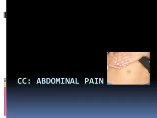
CC: Abdominal pain
A 36-year-old female presents with severe abdominal pain in her right upper quadrant and epigastrium, nausea, vomiting, anorexia, and other symptoms. Learn about differential diagnosis, physical exam findings, and diagnostic tests such as ultrasound and HIDA scan.

CC: Abdominal pain
E N D
Presentation Transcript
HPI • 36 year old female come to the Emergency Department with abdominal pain in her right upper quadrant and epigastrium. The pain is sharp and colicky in nature. She has experienced similar pain in the past but it usually does not last more than a few hours. This pain has been continuous and 8/10 since the company barbeque yesterday afternoon. The pain also radiates to her to her right shoulder and back. Other symptoms include nausea, vomiting, and anorexia. She also complains of subjective fever, sweating and chills.
HPI Past Medical Hx: Family and Social Hx: • Hx irritable bowel syndrome • Hx of fibromyalgia • Hx of laproscopic surgery to remove fibroids 1 week ago • No other past surgical hx • G8P9, all spontaneous vaginal deliveries • No other past surgical history • Mother with hx of HTN, DM and HLD. • Father has hx of obesity, and IBS. • 2 Brothers who are healthy • 1 sister with hx of cholecystitis and cholecystectomy. • Married, lives at home with husband, nine children and 4 dogs. Works at KFC.
DIFFERENTIAL DIAGNOSIS • Biliary colic • Acute pancreatitis. • Appendicitis. • Acute hepatitis. • Peptic ulcer disease. • Fitz-Hugh-Curtis syndrome (perihepatitis caused by gonococcal infection). • Subhepatic or intraabdominal abscess. • Perforated viscus. • Cardiac ischemia. • Black widow spider envenomation • Right-sided pneumonia. • Diseases of the right kidney.
PHYSICAL EXAM VITALS: BP 125/85, T 37.4, R 23, HR 122 GEN: Appears to be in moderate distress secondary to pain. HEENT: Normocephalic, atraumatic (NCAT), pupils equal, round and reactive (PERRL) and extraocular movements intact (EOMI). Moist mucous membranes. Oropharynx clear and patent, no erythema. CV, RESP: Tachycardic, regular rhythm, no murmurs, rubs or gallops. Breath sounds are clear to auscultation bilaterally (CTAB), no crackles or ronchi, and symmetrical chest rise. ABDOMINAL: Soft, non-distended abdomen, voluntary guarding no rebound tenderness. Positive Murphy’s sign. Non-tender McBurney’s point. PELVIC: No bleeding in vaginal vault, no adnexal tenderness, no vaginal discharge or odor. RECTAL: DRE is negative for gross and occult blood. Perianal area is intact without lesions. NEURO: Alert, awake and oriented to person, place and time. (AAOx3). CN II-VII intact. Strength 5/5 and sensation intact over all extremities.
Common Presentation: Classically, patients with cholecystitis present as ill-appearing, febrile and tachycardic. They are often lying still, as acute cholecystitis is associated with local peritoneal inflammation that is aggravated by movement. Abdominal exam often involves voluntary and involuntary guarding. Positive Murphy’s sign is classic in cases of acute cholecystitis. This maneuver entails deep palpation just beneath the liver edge, the patient then inspires deeply, causing the gallbladder to descend toward the examiners hand. In the event of acute cholecystitis the patient will experience increased discomfort and have associated inspiratory arrest. Studies have indicated that sensitivity and specificity for this test are 97 and 48% respectively.
LABORATORY TESTS • CBC: normal • WBC: 16,000, 15 bands • CMP: normal • LFT: AST 50, ALT 65, Alkphos310, tBili <1 • UA: Negative for WBC, RBC, protein, leukocyte esterase and nitrites. • Urine HCG: Negative • What would you like to do next?
Ultrasound SONOGRAPHIC FEATURES • Gallbladder wall thickening >4-5mm or edema (double wall sign) • “Sonographic Murphy’s Sign”, similar to response ilicited by palpation as described previously. Though this is considered more accurate on US, as direct pressure to the GB is confirmed. • Presence of stones in the GB , in the appropriate clinical setting of RUQ pain is suggestive of the diagnosis of cholecystitis, though not diagnostic. • US sensitivity and specificity are 88% and 80% respectively for acute cholecystitis Physical exam is often insufficient to determine specific abdominal viscera as the source of peritoneal inflammation and pain. Ultrasonography can confirm diagnosis. US is usually the first imaging study when cholecystitis is suspected, as it is fast, with minimal risk and low cost in comparison to other imaging studies available.
HIDA Scan • Cholescintigraphy (HIDA scan) is • Cholescintigraphy(HIDA scan) — indicated if the diagnosis remains uncertain following ultrasonography. A nuclear medicine scan using technetium labeled hepatic iminodiacetic acid (HIDA), taken up by the hepatocytes and excreted into bile. Entrance into the gallbladder indicates patency of the cystic duct. HIDA scans will also demonstrate patency of the common bile duct and ampulla. • A positive HIDA scan occurs when the gallbladder does not visualize, due to cystic duct obstruction, most often secondary to edema from acute cholecystitis or an obstructing stone. • Sensitivity and specificity are 97 and 90 percent, respectively. False positives occur in cases of severe liver disease, patients on TPN, obstructive tumors, biliarysphincterotomy, or hyperbilirubinemia. Normal, negative HIDA Positive HIDA
Cholecystitis • Acute cholecystitis refers to a syndrome of right upper quadrant pain, fever, and leukocytosis associated with gallbladder inflammation, often related to gallstone disease. • The pain may radiate to the right shoulder or back. Characteristically, the pain is steady and severe. Associated complaints may include nausea, vomiting, and anorexia. • Acute cholecystitis should be suspected when a patient presenting with the clinical manifestations outlined above is found to have gallstones on an imaging study. Thus, confirmation of the diagnosis must be based upon a combination of physical findings, laboratory studies, and imaging tests . Imaging helps to differentiate from benign biliary colic. • The pain of biliary colic typically reaches a crescendo, and then resolves completely. Pain resolution occurs when the gallbladder relaxes, permitting stones to fall back from the cystic duct. An episode of right upper quadrant pain lasting for more than four to six hours should raise suspicion for acute cholecystitis. As would sx of malaise and fever. • Left untreated, symptoms of cholecystitis may abate within 7 to 10 days, however, complications can occur, including perforation, fistula or infection.
Histological Interpretations Acute cholecystitis results from chemical irritation and inflammation of the obstructed gallbladder. Mucosal phospholipases hydrolyze luminal lecithens to toxic lysolecithins. The normally protective glycoprotein mucus layer is disrupted. This exposes the mucosal epithelium to the direct detergent action of bile salts. Prostaglandins released from GB wall also contribute to mucosal and mural inflammation.
Histological Features Cont’d • The GB is usually enlarged and tense, and often assumes a bright red or blotchy, violaceous to green-black discoloration secondary to subserosal hemorrhages. The serosal covering is frequently layered by fibrin. And possilblysuppurative, coagulated exudate. • In the setting of acute and choroniccholecystitis, see gastric mucin cell metaplasia. Note the conversion from an absorptive columnar cell epithelium (right) to a mucinous epithelium (left).
Treatment • Patients diagnosed with acute cholecystitis require hospital admission for intravenous hydration, correction of electrolyte disorders, and pain control. Patients should be kept fasting and those who are vomiting may need placement of a nasogastric tube. • For patients requiring hospitalization empiric antibiotic therapy is recommended if infection is suspected. • Treatment and timing of definitive therapy depends upon the severity of symptoms and the patient's overall risk of surgery. • Prompt surgical intervention decreases hospital readmission rates and mortality. • High-risk patients, who continue to have severe symptoms and show no appreciable improvement despite one to two days of medical management may require further intervention with gallbladder drainage. • When the cholecystitis has resolved, patients who are surgical candidates should undergo cholecystectomy. • Surgery may also be required when the patient does not improve following percutaneous drainage, which suggests that the gallbladder has already progressed to gangrene. • Patients who stabilize but continue to be at high risk for surgery can be considered for percutaneous gallstone extraction with or without mechanical lithotripsy.
References: • Kumar, Vinay, Abul K. Abbas, Nelson Fausto, Stanley L. Robbins, and RamziS. Cotran. "Chapter 18 Liver and Biliary Tract."Robbins and CotranPathologic Basis of Disease. Philadelphia: Elsevier Saunders, 2005. 882-886. Print. • Zakko, Salam F., MD, FACP, and Nezam H. Afdhal, MD, FRCPI. "Pathogenesis, Clinical Features, and Diagnosis of Acute Cholecystitis." UpToDate. N.p., 5 Oct. 2012. Web. 20 Dec. 2012. <http://www.uptodate.com.proxy.medlib.iupui.edu/co ntents/pathogenesis-clinical-features-and-diagnosis-of-acute- cholecystitis?source=search_result&search=cholecystitis&selected Title=1~150>. • Zakko, Salam F., MD, FACP, Nezam H. Afdhal, MD, FRCPI, and Charles M. Vollmer, Jr, MD. "Treatment of Acute Cholecystitis." Treatment of Acute Cholecystitis. N.p., 25 Sept. 2012. Web. 20 Dec. 2012. <http://www.uptodate.com.proxy.medlib.iupui.edu/contents/treat mentofacutecholecystitis?source=search_result&search=cholecystitis&selectedTitle=2~150>.