Download
1 / 44
681 likes | 3.51k Vues
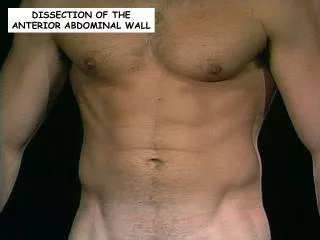
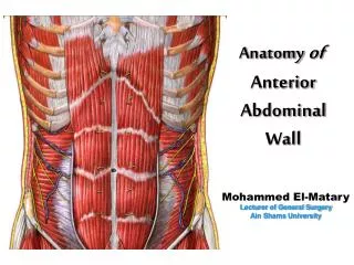
Anatomy of Anterior Abdominal Wall. Mohammed El-Matary Lecturer of General Surgery Ain Shams University. Anterior Abdominal Wall. Layer of anterior abdominal wall: A- Lateral: 1- Skin. 2- Subcutaneous tissue. 3- External oblique muscle.

E N D
Anatomyof Anterior Abdominal Wall Mohammed El-Matary Lecturer of General Surgery Ain Shams University
Anterior Abdominal Wall • Layer of anterior abdominal wall: A- Lateral: 1- Skin. 2- Subcutaneous tissue. 3- External oblique muscle. 4- Internal oblique muscle. 5- Transversus abdominis muscle. 6- Fascia transversalis. 7- Peritoneum.
Anterior Abdominal Wall B- Medial: 1- Skin. 2- Superficial fascia. 3- Anterior wall of rectus sheath. 4- Rectus muscle. 5- Posterior wall of rectus sheath. 6- Peritoneum.
A- Lateral 1- Skin
A- Lateral 2- Subcutaneous tissue
A- Lateral 3- External Oblique m.
A- Lateral 4- Internal Oblique m.
A- Lateral 5- Transversus abdominis m.
A- Lateral 6- Fascia Transversalis m
A- Lateral 7- Peritoneum
Anterior Abdominal Wall • Layer of anterior abdominal wall: B- Medial: 1- Skin. 2- Superficial fascia. 3- Anterior wall of rectus sheath. 4- Rectus muscle. 5- Posterior wall of rectus sheath. 6- Peritoneum.
B- Medial 1- Skin
B- Medial 2- Subcutaneous tissue
B- Medial 3- Ant. Wall of Rectus sheath
B- Medial 4- Rectus Muscle
B- Medial 5- Post. Wall of Rectus sheath
B- Medial 6- Peritoneum
External Oblique Muscle
External Oblique Muscle Origin Fleshy digitations from the lower 8 ribs
External Oblique Muscle Xiphoid Process Insertion The muscle is inserted by fleshy fibers as well as aponeurosis, as follows: A- Fleshy fibers: Outer lip of the iliac crest B- Aponeurosis: • Medial part → linea alba from xiphoid process to symphysis pubis • Lateral part → folded upwards & backwards upon itself to form the inguinal ligament (ASIS → pubic tubercle) Symphysis Pubis
External Oblique Muscle Direction of fibers Downward Forwards Medially
External Oblique Muscle Nerve Supply Intercostal nerves (T7 -T11) & Subcostal nerve (T12)
Internal Oblique Muscle
Internal Oblique Muscle Origin • Anterior 2/3 of the intermediate line of the iliac crest • The lateral 2/3 of the inguinal ligament • Lumbar fascia Insertion • Lower 6 costal cartilages • Xiphoid process • Linea Alba • Pubic crest
Internal Oblique Muscle Direction of fibers Upwards Forwards Medially Nerve Supply T7-T12 Iliohypogastric n. Ilioinguinal n.
Transversus Abdominis Muscle
Transversus Abdominis Muscle 1- Lower 6 intercostal cartilages Origin 4- Lat. 1/3 of inguinal ligament 2- Lumbar Fascia 3- Ant. 2/3 of inner lip of iliac crest
Transversus Abdominis Muscle Insertion 1- Xiphoid Process 3- Linea Alba 2- Pubic Crest
Transversus Abdominis Muscle Direction of fibers Horizontally
Transversus Abdominis Muscle Nerve Supply T7-T12 Iliohypogastric n. Ilioinguinal n.
Rectus Abdominis Muscle
Rectus Abdominis Muscle Insertion Origin 7th, 6th, 5th costal cartilages Xiphoid process From the pubic crest
Rectus Abdominis Muscle Surgical Importance The muscle is divided into segments by tendinous intersections, Which indicate that the muscle arises from a number of myotomes, fused together 1- Segmental nerve supply. 2- Hematoma of rectus m. is localized 2- In paramedian incision displace m. laterally (n. supply comes from lateral)
Pyramidalis Muscle It is a landmark of linea alba intraoperative
Actions of Anterior Abdominal Wall Muscles • They assist in raising the intra-bdominal pressure (so, they help in vomiting, cough, delivery, etc….) • Keep the abdominal viscera in position. • Rectus abdominis flexes the trunk, while the 2 oblique muscles bend the trunk laterally. • Act as accessory expiratory muscles. • Lower midline & paramedian incisions.
Rectus Sheath Linea Alba Medially Arcuate Line Linea Semilunaris Laterally
Rectus Sheath Above Arcuate Line External Oblique Ant. Layer of Rectus Sheath Internal Oblique Transversus Abdominis Rectus Abdominis SKIN Falciform Ligament Peritoneum Transverslais Fascia Post. Layer of Rectus Sheath
Rectus Sheath Below Arcuate Line External Oblique Ant. Layer of Rectus Sheath Internal Oblique Transversus Abdominis Rectus Abdominis SKIN Transverslais Fascia Peritoneum Urachus in Median Umbilical Fold Medial Umbilical Ligament
Arterial Supply of Anterior Abdominal Wall
- I -Internal Mammary a. - II - Descending Aorta Superior epigastric a. 10th, 11th intercostal a. Subcostal a. - III -External Iliac a. Deep circumflex iliac a. Inferior epigastric a.
Thanks You Mohamed Noman