Download

1 / 34
440 likes | 1.49k Vues
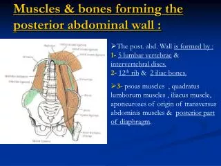
Posterior abdominal wall. Posterior abdominal wall. Structures of post. Abdominal wall: - 5 lumbar vertebra & their intervertebral disc - 12 th ribs - Upper part of bony pelvis - Muscles - psoas major - psoas minor - Quadratus lumborum

E N D
Posterior abdominal wall Structures of post. Abdominal wall: - 5 lumbar vertebra & their intervertebral disc - 12th ribs - Upper part of bony pelvis - Muscles - psoas major - psoas minor - Quadratuslumborum - Aponeurosis of transversusabdominis muscles iliacus muscle lie in the iliac fossa
Muscles of post.abdominal wall Psoas major • Origin: body & transverse process of lumbar vertebra & intervertebral disc • Ins :Lesser trochanter of femur • N.S: nerve plexus(T12,L1,L2.L3) • Action: flexion of hip & thigh Quadratuslumborum • Origin: Iliolumbarlig.& iliac crest • Ins: 12th rib • N.S: nerve plexus(T12,L1,L2.L3 • Action: fix or depresses 12th rib during respiration & lateral flexion of the trunk Iliacus muscle • Origin: iliac fossa • Ins: Lesser trochanter of femur • N.S: femoral nerve • Action: Lateral flexion of hip & thigh for lying position
Iliolumbar ligament The Iliolumbar ligament is a strong ligamentpassing from the tip of the transverse process of the fifth lumbar vertebra to the posterior part 1/3 of the inner lip of the iliac crest
Arteries on the Posterior Abdominal Wall Aorta Location and Description • The aorta enters the abdomen through the aortic opening of the diaphragm in front of the 12th thoracic vertebra . • It descends behind the peritoneum on the anterior surface of the bodies of the lumbar vertebrae. • End: At the level of the fourth lumbar vertebra, it divides into two common iliac arteries . Relation Ant: - Pancreas - 3rd part of d.d - Coils of small intestine - Crossed by Lt.renal vein On its right side : - The inferior vena cava - The cisternachyli - The beginning of the azygos vein. On its left side - The left sympathetic trunk.
Branches of abd. Aorta • Single branches: - 3 front & I back Front: - The celiac artery - Superior mesenteric artery - Inferior mesenteric artery Back: - Median sacral artery
Abdominal Aorta……cont Pairs branches: 1 front, 4back & 3 side of aorta 1- 1 front testicular or ovarian artery at level L2 2- 4 back lumbar arteies 3- 3 side of aorta - Inferior phrenic . a - Middle suprarenal . a - Renal . a
Abdominal aorta…cont • Celiac Artery at level L1 • The celiac artery or trunk is very short and arises from the commencement of the abdominal aorta at the level of the 12th thoracic vertebra. It is surrounded by the celiac plexus and lies behind the lesser sac of peritoneum. • It has three terminal branches: 1- The left gastric - The small left gastric artery runs to the cardiac end of the stomach, gives off a few esophageal branches, then turns to the right along the lesser curvature of the stomach. It anastomosis with the right gastric artery 2- Splenic Artery - Pancreatic branches • The left gastroepiploic artery • The short gastric arteries fundus 3- hepatic arteries • The right gastric artery arises from the hepatic artery at the upper border of the pylorus and runs to the left in the lesser omentum along the lesser curvature of the stomach. It anastomosis with the left gastric artery . • The gastroduodenal artery : - It divides into the right gastroepiploic artery that runs along the greater curvature of the stomach between the layers of the greater omentum and the superior pancreaticoduodenal artery that descends between the second part of the duodenum and the head of the pancreas . • The right and left hepatic arteries enter the portahepatis. The right hepatic artery usually gives off the cystic artery, which runs to the neck of the gallbladder
Abdominal aorta……cont • Superior mesenteric Artery at level L 2 • The inferior pancreaticoduodenal artery • The middle colic artery runs forward in the transverse mesocolon to supply the transverse colon and divides into right and left branches. • The right colic artery is often a branch of the ileocolic artery. It passes to the right to supply the ascending colon and divides into ascending and descending branches. • The ileocolic artery passes downward and to the right. It gives rise to a superior branch that anastomoses with the right colic artery and an inferior branch that anastomoses with the end of the superior mesenteric artery. The inferior branch gives rise to the anterior and posterior Cecal arteries; the appendicular artery is a branch of the posterior Cecal artery . • The jejunal and ileal branches are 12 to 15 in number and arise from the left side of the superior mesenteric artery . Each artery divides into two vessels, which unite with adjacent branches to form a series of arcades. Branches from the arcades divide and unite to form a second, third, and fourth series of arcades. Fewer arcades supply the jejunum than supply the ileum. From the terminal arcades, small straight vessels supply the intestine
Abdominal aorta….cont • Inferior Mesenteric Artery at level L3 Branches • The left colic artery runs upward and to the left and supplies the distal third of the transverse colon, the left colic flexure, and the upper part of the descending colon. It divides into ascending and descending branches. • The sigmoid arteries are two or three in number and supply the descending and sigmoid colon. • The superior rectal artery is a continuation of the inferior mesenteric artery as it crosses the left common iliac artery. It descends into the pelvis behind the rectum. The artery supplies the rectum and upper half of the anal canal and anastomoses with the middle rectal and inferior rectal arteries
Marginal Artery The anastomosis of the colic arteries around the concave margin of the large intestine forms a single arterial trunk called the marginal artery. This begins at the ileocecal junction, where it anastomoses with the ileal branches of the superior mesenteric artery, and it ends where it anastomoses less freely with the superior rectal artery
Common Iliac Arteries The right and left common iliac arteries are the terminal branches of the abdominal aorta. They arise at the level of the fourth lumbar vertebra and run downward and laterally along At medial border of the psoas muscle . Each artery ends in front of the sacroiliac joint by dividing into the external and internal iliac arteries. At the bifurcation, the common iliac artery on each side is crossed anteriorly by the ureter .
External Iliac Artery runs along the medial border of the psoas, following the pelvic brim . It gives off the inferior epigastric and deep circumflex iliac branches . The artery enters the thigh by passing under the inguinal ligament to become the femoral artery.
Branches of Ex. iliac artery 1- The inferior epigastric - artery arises just above the inguinal ligament. - It passes upward and medially along the medial margin of the deep inguinal ring and enters the rectus sheath behind the rectus abdominis muscle. 2- The deep circumflex iliac - artery arises close to the inferior epigastric artery . - It ascends laterally to the anterior superior iliac spine and the iliac crest, supplying the muscles of the anterior abdominal wall.
Internal Iliac Artery • The internal iliac artery passes down into the pelvis in front of the sacroiliac joint • Posterior Iliolumbar artery • Posterior Lateral sacral arteries • Posterior Superior gluteal artery - greater sciatic foramen • Anterior Obturator artery (occasionally from inferior epigastric artery) - obturator canal • Anterior Inferior gluteal artery - greater sciatic foramen • Anterior Umbilical arterysuperior vesical artery (usually, but sometimes it branches directly from anterior trunk) medial umbilical ligament • Anterior Uterine artery (females) or deferential artery (males) superior and vaginal branches uterus, vas deferens • Anterior Vaginal artery (females, can also arise from uterine artery) - vagina • Anterior inferior vesical artery - urinary bladder • Anterior Middle rectal artery - rectum • Anterior Internal pudendal artery
Veins on the Posterior Abdominal Wall Inferior Vena Cava - Location and Description • The inferior vena cava conveys most of the blood from the body below the diaphragm to the right atrium of the heart. • It is formed by the union of the common iliac veins behind the right common iliac artery at the level of the fifth lumbar vertebra . • It ascends on the right side of the aorta, pierces the central tendon of the diaphragm • Ascends then separated from the aorta by Rt .crus of the diaphragm • Ends at the level of the eighth thoracic vertebra, and drains into the right atrium of the heart. • The right sympathetic trunk lies behind its right margin and the right ureter lies close to its right border. The entrance into the lesser sac separates the inferior vena cava from the portal vein .
Relations of I.V.C • Anterior - Coils of small intestine - 3rd part & 1st part of d.d - Head of pancreas & C.B.D - Related to foramen of Winslow - Portal vein - Lies in deep groove of liver
Tributaries of I.V.C • The inferior vena cava has the following tributaries • Two anterior visceral tributaries: the hepatic veins • Three lateral visceral tributaries: the right suprarenal vein (the left vein drains into the left renal vein), renal veins, and right testicular or ovarian vein (the left vein drains into the left renal vein) • Five lateral abdominal wall tributaries: the inferior phrenic vein and four lumbar veins • Three veins of origin: two common iliac veins and the median sacral vein
Inferior Mesenteric Vein • The inferior mesenteric vein is a tributary of the portal circulation. It begins halfway down the anal canal as the superior rectal vein . It passes up the posterior abdominal wall on the left side of the inferior mesenteric artery and the duodenojejunal flexure and joins the splenic vein behind the pancreas. It receives tributaries that correspond to the branches of the artery. • The splenic vein • It is a tributary of the portal circulation. It begins at the hilum of the spleen by the union of several veins and is then joined by the short gastric and the left gastroepiploic veins. It passes to the right within the splenicorenal ligament and runs behind the pancreas. It joins the superior mesenteric vein behind the neck of the pancreas to form the portal vein. It is joined by veins from the pancreas and the inferior mesenteric vein. • Superior Mesenteric Vein • The superior mesenteric vein is a tributary of the portal circulation. It begins at the ileocecal junction and runs upward on the posterior abdominal wall within the root of the mesentery of the small intestine and on the right side of the superior mesenteric artery. It passes in front of the third part of the duodenum and behind the neck of the pancreas, where it joins the splenic vein to form the portal vein. It receives tributaries that correspond to the branches of the superior mesenteric artery and also receives the inferior pancreaticoduodenal vein and the right gastroepiploic vein .
Portal Vein • The portal vein drains blood from the abdominal part of the gastrointestinal tract from the lower third of the esophagus to halfway down the anal canal; it also drains blood from the spleen, pancreas, and gallbladder. • The portal vein enters the liver and breaks up into sinusoids, from which blood passes into the hepatic veins that join the inferior vena cava. • It is about 2 in. (5 cm) long and is formed behind the neck of the pancreas by the union of the superior mesenteric and splenic veins. • It ascends to the right, behind the first part of the duodenum, and enters the lesser omentum . • It then runs upward in front of the opening into the lesser sac to the portahepatis, where it divides into right and left terminal branches • The portal circulation begins as a capillary plexus in the organs it drains and ends by emptying its blood into sinusoids within the liver.
. Tributaries of the Portal Vein • The tributaries of the portal vein are the splenic vein, superior mesenteric vein, left gastric vein, right gastric vein, and cystic veins. • Splenic vein: This vein leaves the hilum of the spleen and passes to the right in the splenicorenal ligament. It unites with the superior mesenteric vein behind the neck of the pancreas to form the portal vein . It receives the short gastric, left gastroepiploic, inferior mesenteric, and pancreatic veins. • Inferior mesenteric vein: This vein ascends on the posterior abdominal wall and joins the splenic vein behind the body of the pancreas. It receives the superior rectal veins, the sigmoid veins, and the left colic vein. • Superior mesenteric vein: This vein ascends in the root of the mesentery of the small intestine. It passes in front of the third part of the duodenum and joins the splenic vein behind the neck of the pancreas . It receives the jejunal, ileal, ileocolic, right colic, middle colic, inferior pancreaticoduodenal, and right gastroepiploic veins. • Left gastric vein: This vein drains the left portion of the lesser curvature of the stomach and the distal part of the esophagus. It opens directly into the portal vein • Right gastric vein: This vein drains the right portion of the lesser curvature of the stomach and drains directly into the portal vein . • Cystic veins: These veins either drain the gallbladder directly into the liver or join the portal vein.
Portal systemic anastomosis A portacavalanastomosis (also known as portal systemic anastomosis or portal caval system) is a specific type of anastomosis that occurs between the veins of portal circulation and those of systemic circulation. The lower end of esophagus is one of the important sites for the portosystemicanastomosis . In portal hypertension as in the case of cirrhosis of liver the anastomosis opens and forms venous dilatation called esophageal varices. Their rupture causes severe and dangerous haematesis (hematemesis).
Portal systemic anastomosis……cont Causes • Liver diseases Cirrhosis, fibrosis ( bilharzial) • Valvular diseases of the heart • Congenital patent
Lymphatics on the Posterior Abdominal Wall • Lymph Nodes • The lymph nodes are closely related to the aorta and form a preaortic and a right and left lateral aortic (Para-aortic or lumbar) chain . • The preaortic lymph nodes • lie around the origins of the celiac, superior mesenteric, and inferior mesenteric arteries and are referred to as the celiac, superior mesenteric, and inferior mesenteric lymph nodes, respectively. • They drain the lymph from the gastrointestinal tract, extending from the lower one third of the esophagus to halfway down the anal canal, and from the spleen, pancreas, gallbladder, and greater part of the liver. • The efferent lymph vessels form the large intestinal trunk. • The lateral aortic (para-aortic or lumbar) lymph nodes • drain lymph from the kidneys and suprarenals; from the testes in the male and from the ovaries, uterine tubes, and fundus of the uterus in the female; from the deep lymph vessels of the abdominal walls; and from the common iliac nodes. • The efferent lymph vessels form the right and left lumbar trunk • The thoracic duct commences in the abdomen as an elongated lymph sac, the cisternachyli. This lies just below the diaphragm in front of the first two lumbar vertebrae and on the right side of the aorta .
The cisternachyli • The right and left lumbar trunks under the diaphragm on the side of the aorta • Receives lymph from - The intestinal trunk - Some small lymph vessels that descend from the lower part of the thorax. - Rt & Lt vessels from lower thorax