Download
1 / 15
210 likes | 1.28k Vues
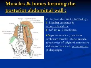
Muscles & bones forming the posterior abdominal wall :. The post. abd. Wall is formed by : 1- 5 lumbar vertebrae & intervertebral discs. 2- 12 th rib & 2 iliac bones.

E N D
Muscles & bones forming the posterior abdominal wall : • The post. abd. Wall is formed by :1-5 lumbar vertebrae & intervertebral discs.2-12th rib & 2 iliac bones. • 3- psoas muscles , quadratus lumborum muscles , iliacus muscle, aponeuroses of origin of transversus abdominis muscles & posterior part of diaphragm.
Psoas Major muscle : • Origin : transverse processes , sides of bodies & intervertebral discs of 12th thoracic & the 5lumbar vertebrae. • Insertion : with iliacus into the lesser trochanter of femur, by passing behind inguinal ligamen. • Nerve supply : lumbar plexus(L1,2,3) • Action :flexes thigh on trunk at hip joint & if thigh is fixed , it flexes the trunk onthigh , as in sitting up from lying position. • The psoas is enclosed in a fibrous sheath derived from the lumbar fascia, this sheath is thickened above forming medial arcuateligament.
Psoas Minor Muscle : • It is a small unimportant muscle, absent in 40% of people, if it is present it lies in front of psoas major • Origin : adjacent sides of bodies of T12 & L1 vertebrae & disc between them. • Insertion : iliopectineal eminence & pectineal line. • N.Supply : ventral ramus of L1 N. • Action : Flexion of lumbar part of V.column.
Quadratus Lumborum muscle : • Origin : iliolumbar ligament , iliac crest , tips of transverse processes of lower 2-3 lumbar vertebrae. • Insertion : lower border of 12th rib & upper 4 lumbar vertebrae. • Nerve supply : lumbar plexus(L1,2,3,4). • Action :fixation of 12th rib to help contraction of diaphragm during deepinspiration– depresses 12th rib during forced expiration – lateral flexion of vertebral column on the same side. • It is covered by lumbar fascia , which is thickened above forming lateral arcuate ligament , and below forming iliolumbar ligament.
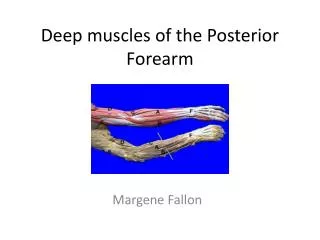
Iliacus muscle : • Origin : iliac fossa • Insertion : with the psoas major tendon.... into the lesser trochanter of femur. The combined muscles are called iliopsoas. • Nerve supply : femoral N. (branch of lumbar plexus). • Action : 1-iliopsoas flexes the thigh ontrunk at hip joint , or if the thigh is fixed… 2-.It flexes the trunk onthigh. (as psoas major ).
Fascial lining of the abdominal walls • The abdominal walls are lined by one continuous layer of C.T.that lies between parietal peritoneum & abdominal muscles • It is continuous below with a pelvic fascia lining pelvic walls. • Its name is according to structure it overlies. Diaphragmatic fascia covers undersurface of diaphragm, transversalis fascia lines transversus abdominis, psoas fascia covers psoas muscle, Quadratus lumborum fascia covers Q.L.muscle & iliaca fascia covers iliacus muscle. • Abdominal blood & lymph vessels lie within fascial lining, while nerves lie outside the fascia as in femoral sheath.
Thoraco-lumbar Fascia : • It is a deep fascia which covers and encloses the muscles of the back. • Lumbar part of deep fascia lies in interval between iliac crest & 12th rib. • Laterally : it gives origin to : 1-middle Fs.of transversus abdominis. 2-upper Fs. of internal oblique. • Medially : it splits into 3 lamellae or layers : Anterior + middle layers, extend superiorly to last rib & inferiorly to iliac crest, & (enclose Q.L.muscle). Posterior layer extends upwards to thorax & neck & (enclose erector spinae muscle).
Posterior layer : -it covers back of erector spinae ms –it extends from sacrum to neck. -in the lumbar region , it is very thick & attached to lumbar spines. • Middle layer : -it covers back of Q.L.muscle. -Medially, it attached to transverse processes of lumbar vertebrae. -Laterally it unites with post. layer to give origin toint.oblique ms. & with anterior layer to give origin totransversus abdominis ms. lumbar Fascia • Anterior layer : -it lies infront ofQ.L.ms. -medially, it is attached to the lumbar transvese processes. -laterally : it fuses with middle layer to give origin to middle Fs. of transversus abdominis ms. • Q.L.muscle is enclosed between anterior & middle layers of thoraco-lumbar fascia.
Fascia of psoas & iliacus • It is the fascial sheet covering the ventral aspects of psoas & iliacus. • Above iliac crest : -it covers psoas ms. Only. - Medially, it is attached to lumbar vertebrae & fused laterally with anterior layer of thoraco-lumbar fascia • Below iliac crest : -it is called fascia iliaca as it coverspsoas & iliacus.
Fascia iliaca • Below iliac crest : Medially : it is attached to pelvicbrim(sacral promontory + pectineal line + sympysis pubis). Laterally : it is attached to iliac crest. • -inferiorly , Behind inguinal lig. :- its lateral part : it fuses with fascia transversalis behind lateral part of inguinal lig, -but its medial part descends to the thigh behind femoral vessels to form post. wall of femoralsheath.
Iliopsoas Fascia and Tuberculosis : • T.B. disease of thoraco-lmbarregion of vertebral column. results in destruction of vertebral bodies with possible extension of pus laterally under the psoas & iliac fascia to appear as a swelling below the inguinal ligament ( may be mistaken for femoral hernia).
Relations of Psoas Major muscle : • Inside its substances, Lumbar plexus is formed.(L1,2,3,4). Anteriorly : it is covered by psoas fascia, psoas minor and is related to: kidney,ureter,renal vs., gonadal vs.,genitofemoral N.Posteriorly : lumbar transverse processes + medial edge of Q.L. • Medially : lumbar vertebral bodies, lumbar vs., & symp.trunk. • Laterally : these Ns. emerge from its lateral border : iliohypogastric N.,ilioinguinal N.,lat.cut.N.of thigh & femoral N., which lies in deep groove between psoas major & iliacus.
Relations of Quadratus lumborum muscle : • Anteriorly : 1- it is covered by Q.L.fascia . 2-it is closed between anterior & middle layers of thoracolumbar fascia. 3-subcostal vs.&N., iliohypogastric & ilioinguinal nerves. 4-psoas major & minor. 5-colon & kidney. • Posteriorly : erector spinae muscle.
Relations of Iliacus muscle : • Anteriorly : 1-fascia iliaca. 2-lateral cut.N. of thigh. 3-Rt.muscle is related to caecum, while Left muscle is related to descending & pelvic colon. • Posteriorly : iliac fossa. • Medially : Psoas major + femoral N. ( in deep groove between psoas & iliacus ).