Anterior abdominal wall
Anterior abdominal wall. THE ABDOMINOPELVIC CAVITY. largest effectively continuous visceral cavity of the body. provide multiple vital functions support and protection of the digestive and urinary tracts and internal reproductive organs and their associated neurovascular supplies.


Anterior abdominal wall
E N D
Presentation Transcript
THE ABDOMINOPELVIC CAVITY • largest effectively continuous visceral cavity of the body. • provide multiple vital functions • support and protection of the digestive and urinary tracts and internal reproductive organs and their associated neurovascular supplies
transmission of the neurovascular supply to and from the thorax and the lower limb • provision of support and attachment to the external genitalia and access to and from the internal reproductive and urinary organs
provision of accessory muscles of physiological actions such as respiration, defecation, and micturition • support for the spinal column in weight bearing and movement.
MUSCULOSKELETAL FRAMEWORK • five lumbar vertebrae and their intervening intervertebral discs (lying in the posterior midline) • three layers of skeletal muscles (transversusabdominis, internal oblique and external oblique) with associated fasciae and skin (lying lateral and anterolateral
a single muscular layer (rectus abdominis) with its associated fascial coverings (lying anterior) • the bony ‘bowl' formed by the walls of the true and false pelvis (ilium, ischium and pubis on each side
the muscles of the pelvic floor and perineum (lying inferiorly) • the diaphragm (lying superiorly)
Bony protection • the pelvis (true and false) • anterolateral portions of the lower six ribs and their cartilages even though these structures are technically part of the thoracic wall.
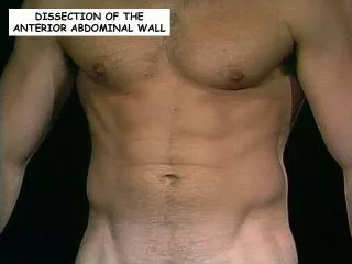
Between these two zones, the anterolateral abdominal wall is entirely musculofascial but of sufficient thickness and strength that it provides adequate protection for the viscera such that even direct blows can be resisted.
The abdominal wall and retroperitoneal structures play an important role in the function of the spinal column in both movement of the thorax in relation to the pelvis and in aiding support of the spine in weight bearing.
The anterolateral muscles provide assistance with flexion and rotation of the thorax in relation to the pelvis (or vice versa if the thorax is fixed).
Communications between thoracic and abdominal cavities • Inferior vena cava crosses between its through the caval opening of the diaphragm • Oesophagus passing inferiorly through the oesophageal opening of the diaphragm • Aorta between posterior to the median arcuate ligament of the diaphragm
4. Lymphatics of the abdomen draining upwards to the thorax via the thoracic duct, peri-cavallymphatics and small vessels draining directly through and at the peripheral insertions of the diaphragm 5. Azygos and hemiazygos veins ascending into the thoracic azygos system
6. the autonomic nervous system, both sympathetic and parasympathetic via the various diaphragmatic openings, and directly through the substance of the diaphragm itself