Download
1 / 40
490 likes | 1.65k Vues
Abdomen 1. 2.1 Surface anatomy 2.2 Anterior abdominal wall. Albert van Schoor GNK 288 (SA4 Anatomy dissection). 2.1.1 Surface anatomy.

E N D
Abdomen 1 2.1 Surface anatomy2.2 Anterior abdominal wall Albert van Schoor GNK 288 (SA4 Anatomy dissection)
2.1.1 Surface anatomy • Identify and name the bony landmarks of the abdomen which are palpable on abdominal examination and state if possible their corresponding vertebral heights on the cadaver, yourself and on a radiograph • Schematically illustrate and discuss the nine abdominal regions and list which organs lie approximately in each region
2.1.1 Surface anatomy • Identify and name in which of the nine surface anatomical regions you would expect to feel tenderness in appendicitis, cholecystitis, gastritis and cystitis • Identify other surface anatomy lines e.g. transpyloric line, transumbilical line, linea alba and linea semilunaris. You should be able to say how these lines are formed and to discuss the intra-abdominal events occurring on the transpyloric line
2.1.1 Surface Anatomy Referred pain
Transpyloric plane Hilum of the kidneys Pylorus of the stomach Body of pancreas Fundus of the gall bladder 2.1.1 Surface Anatomy
2.1.1 Surface anatomy • Schematically illustrate and discuss the surface anatomy of the kidneys, ureters and spleen on the posterior abdominal wall [2.5, 2.8] • Discuss and identify the surface anatomy of the liver [2.4] • Briefly discuss the surface anatomy of the diaphragm and the vertebral heights of its three major orifices [2.9] • Identify the various dermatomes of the anterior abdominal wall • Indicate whether the bladder is an abdominal organ or not. Explain
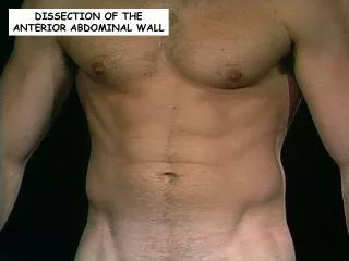
2.2 Anterior abdominal wall 2.2.1 Skin and superficial fascia 2.2.2 Muscles 2.2.3 Transversalis fascia 2.2.4 Peritoneum 2.2.5 Rectus sheath 2.2.6 Inguinal area 2.2.7 Osteology
2.2.1 Skin and superficial fascia • Identify the fatty superficial layer (Camper's fascia) • Identify and shortly discuss the membranous superficial layer (Scarpa's fascia) according to its distribution and borders. State what this fascia is called inferior to the superficial inguinal ring
2.2.2 Muscles • Identify and discuss the three major abdominal muscles as follows: • Major attachments to the following points: Linea alba, crista iliaca and inguinal ligament where applicable, • Direction of fibres, • Nerve supply and • Function • Identify the neurovascular plane • Identify the ilio-inguinal and iliohypogastric nerves. Also state their origin and area of supply
2.2.2 Muscles External oblique Table
2.2.2 Muscles Internal oblique
2.2.2 Muscles Transverse abdominis
2.2.2 Muscles Neurovascular plane Ant Post
2.2.3 Transversalis fascia • Identify the transversalis fascia
2.2.4 Peritoneum • Identify the folds and associated underlying structures of the peritoneum on the posterior aspect of the anterior abdominal wall: [2.3.3] • Plica umbilicalis mediana, • Plica umbilicalis medialis, • Plica umbilicalis lateralis, • Falciform ligament
2.2.5 Rectus sheath • Identify the rectus sheath and how it is formed on the following levels: • Superior to the arcuate line • Inferior to the arcuate line • Identify and briefly discuss rectus abdominis as follows: • major attachments, • nerve supply and • function • Name and identify the structures on the posteriorwall of the rectus sheath
2.2.5 Rectus sheath Rectus abdominis
2.2.6 Inguinal area • Identify and briefly discuss the inguinal canal as follows: • Surface anatomy, • Borders, • Openings
2.2.6 Inguinal area Inguinal canalSurface anatomy
2.2.6 Inguinal area Indirect inguinal hernia
2.2.6 Inguinal area Inguinal canalBorders 491-8
2.2.6 Inguinal area • Know the positions of the superficial and deep inguinal rings and femoral canal. • Identify the inguinal ligament and the structures posterior to it from lateral to medial.
2.2.6 Inguinal area Femoral canal Ant Post
2.2.6 Inguinal area Femoral hernia
2.2.6 Inguinal area N.A.V.E.L Contents • N.A.V.E.L (lat. - med.) • Femoral nerve • Femoral artery • Femoral vein • Empty space (femoral canal) • Lacunar ligament N A V E L
2.2.6 Inguinal area • List the contents of the spermatic cord. Compare the content in males and females • Identify the inferior epigastric artery and its relation to the deep inguinal ring • Identify and list the borders of the inguinal triangle (Hesselbach's triangle) • Name and identify the inguinal falx (conjoint tendon)
2.2.6 Inguinal area Spermatic cord • 3 Fascia layers • External spermatic fascia • Cremasteric fascia • Internal speratic fascia • 3 Arteries • Testicular artery • Cremasteric artery • Artery to ductus deferens • 3 Nerves • Genito-femoral nerve • Ilio-inguinal nerve • Sympathetic autonomic plexus • 3 Other structures • Lymphatic vessels • Ductus deferens • Pampiniform venous plexus
2.2.6 Inguinal area Spermatic cord
2.2.6 Inguinal area Hesselbach’s triangle
2.2.6 Inguinal area Direct inguinal hernia
2.2.6 Inguinal area Conjoint tendon
2.2.7 Osteology • Identify the following bony points of the os coxa: • Anterior superior iliac spine (ASIS) • Anterior inferior iliac spine (AIIS) • Crista iliaca • Posterior superior iliac spine (PIIS) • Pubic tubercle • Pubic crest • Symphysis pubis