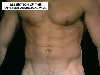
Anterior abdominal wall
Anterior abdominal wall. Layers of Anterior Abdominal Wall. In human anatomy , the layers of the abdominal wall are (from superficial to deep): Skin Fascia Camper's fascia - fatty superficial layer. Scarpa's fascia - deep fibrous layer. Muscle -Rectus abdominis


Anterior abdominal wall
E N D
Presentation Transcript
Layers of Anterior Abdominal Wall • In human anatomy, the layers of the abdominal wall are (from superficial to deep): • Skin • Fascia • Camper's fascia - fatty superficial layer. • Scarpa's fascia - deep fibrous layer. • Muscle -Rectus abdominis -External oblique muscle -Internal oblique muscle -Transverse abdominal muscle Pyramidalis muscle Cremasteric • Fascia transversalis • Parital Peritoneum
Fascia of Ant. Abdominal Wall 1- there is no deep fascia 2-the superficial fascia is formed of a single layer above the ambilicus , but below the ambilicus it differentiated into 2 layers: a- superficial fatty layer (Camper’s fascia) - contains variable amount of fat . b- deep membranes layer (Scarpa’s fascia) -continuous with the superficil perineal fascia (colle’s fascia)
Linea Alba An extensive aponeurosis which extend from the xiphoid process to the symphsis pubis . Linea Semilunaris the lateral margin of rectus abdominis is marked by a line on the anterior abdominal wall called linea semilunaris
Muscles of the anterior abdominal wall Classified into 2 groups: 1-paramedian muscles -Rectus abdominis muscle - Pyramidalis muscle 2- anterolateral flat muscles -external abdominal oblique m. -internal abdominal oblique m. -transversus abdominis m.
Abdominal external oblique muscle • the largest and the most superficial of the three flat muscles of the lateral anterior abdomen.
Origin : lower 8 ribs (outer surface) Direction of the fibers : downwards , forwards & medialy Insertion : -iliac crest ( anterior half of the outer lip) -anterior superior iliac spine -pubic tubercle -pubic crest -linea alba ………………………….
Inguinal ligament -It is the infolded lower border of the external oblique m. -The inguinal ligament runs from the anterior superior iliac spine to the pubic tubercle.
Abdominal internal oblique muscle Origin : 1- iliac crest (ant. 2/3 of its intermediate line) 2- inguinal ligament ( lateral 2/3 of its inner surface). 3- lumbar fascia . Direction of the fibers : upwards , forwards and medialy Insertion : 1- linea alba 2- lower 4 or 5 ribs 3- the lower fibers forms an arched fibers called the conjoint tendon.
Conjoint tendon -Formed by the lower arched fibers of both internal oblique and transversus abdominis muscles where the fiberous pass directly from origin to insertion -Function : The contraction of the conjoint tenden leads to closure of the inguinal canal (shutter mechanism ) -So , it prevent passage of intestine through the canal (Indirect inguinal hernia) .
Transversus abdominis muscle Origin : -the lateral third of the inner surface of inguinal ligament -anterior 2/3 of the medial lip of the iliac crest -the inner surfaces of the cartilages of the lower 8 ribs -the thoracolumbarl fascia. Direction of the fibers : -horizontaly . Insertion: a- linea alba b-the lower fibers form an arched fibers together with the lower fibers of internal oblique muscle (conjoint tendon)
Rectus abdominus m. Site : it lies in the middle part of the abdominal muscle layer inside the tendinous sheath called rectus sheath. Origin : symphysis pupis and pubic crest. Insetion: 5th, 6th & 7th costal cartilages- -The muscle is divided by 3 tendinous intersections as follows: -These tendentious intersections are present on the anterior surface of the muscle and indicate that the muscle arises from different myotomes .
Action : -flexion of the trunk . Surface anatomy : the lateral margin of rectus abdominis is marked by aline on the anterior abdominal wall called linea semilunaris .
Cremastric muscle: • It is formed by loops of muscle fibres from the lower part of the internal oblique • it extends down through the spermatic cord & return behind it to be attached to the pupic tubercle • its n. supply is the genital branch of the genitofemoral n. • action: • elevation of testis upwards in the cold weather. • Cremastric reflex: • Scratching of the upper & medial parts of the thigh (L1) leads to elevation of the testis.
Pyramidalis muscle : origin : pubic crest and symphysis pubis insertion : into the lower part of the linea alba nerve supply : subcostal nerve action : it stretches the linea alba
Action of the anterior abdominal wall muscles 1-support the viscera and keep them in their position 2-contract to increase the intra-abdominal pressure during urination and defection (mainly the trunsversus abdominis). 3-expiratory action : -the abdominal muscles contract during expiration and relax during inspiraion . 4-movement of the trunk : a-contraction of rectus abdominis muscle ……..forward flexion. b-contraction of one side of the oblique muscles ……….lateral flexion . c. combined action of external oblique with the oppisite internal oblique ……rotation of the trunk . 5- elevation of the testis………..by the cremastric m .
Inguinal canal • Definition: It is an oblique inter-muscular canal (4cm) above the medial part of the inguinal ligament. • Openings: -it starts at the deep inguinal ring and ends at the superfascial inguinal ring.
a-Superfascial Inguinal Ring Site: it is a triangular or inverted V shaped opening in the external oblique aponeurosis. its margins give the external spermatic fascia to cover the spermatic cord. Surface anatomy: It lies just above the pupic tubercle. Structures passing through it: -illio-inguinal nerve. -Spermatic cord ( in male) or round ligament ( in female)
B -Deep Inguinal Ring Site: It is a small opening in the lower part of fascia transversalis. Its margins give the internal spermatic fascia to cover the spermatic cord. Surface anatomy: - It lies ½ an inch above the mid-point of the inguinal ligament. Structures passing through it: -Spermatic cord (in male) or round ligament ( in female).
Arteries of the Anterior Abdominal wallA- Above the umbilicus 1-superior epigastric artery 2-musculo-phrenic artery 3-lower two posterior intercostal and subcostal arteries
B- Below the umbilicus 1-Inferior epigastric artery 2-Deep circumflex iliac artery 3-Superficial branches of femoral artery
Veins of Anterior Abdominal Wall The anterior abdominal wall is drained by veins corresponding to the arteries
Nerves of the Anterior Abdominal Wall 1-Lower five intercostal and subcostal nerves 2-Illio-hypogastric and illio-inguinal nerves …………………..
Lymphatics of Anterior Abdominal Wall A-Above the umbilicus into pectoral lymph nodes B- Below the umbilicus into superficial inguinal lymph nodes ……………