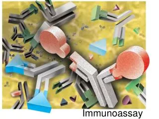
Labeled Immunoassay
Labeled Immunoassay. Lab. 2. An immunoassay is a test that uses antibody and antigen complexes as a mean of generating a result Immuno refers to an immune response that causes the body to generate antibodies, and assay refers to a test

Labeled Immunoassay
E N D
Presentation Transcript
Labeled Immunoassay Lab. 2
An immunoassay is a test that uses antibody and antigen complexes as a mean of generating a result • Immunorefers to an immune response that causes the body to generate antibodies, • and assayrefers to a test • An antibody-antigen complex is also known as an immuno-complex • The assay takes advantage of the specific binding of an antibody to its antigen • The antibodies used must have a high affinity for the antigen Immunoassay
Immunoassays derive their unique specificity, sensitivity, and flexibility from three important properties of antibodies: • Their ability to bind to an extremely wide range of natural and man-made chemicals, biomolecules, cells, and viruses • Exceptional specificity for the substance to which each antibody binds • The strength of the binding between an antibody and its target Immunoassays
Both the presence of antigen or antibodycan be measured • For measuring hormones such as insulin, the insulin acts as the antigen • when detecting infection the presence of antibody against the pathogen is measured • For numerical results the response of the objectbeing measured must be compared to standards of a known concentration • This is usually done through the plotting of a standard curve on a graph paper, and then the quantity of the unknown is found from the curve Immunoassay
History • In the year 1959, Drs. Rosalyn Yalow & SolomanBerson invented the radioimmunoassay (RIA) • Applied the use of radioisotopes in the measurement of insulin • The RIA is the predecessor of modern immunoassays Dr. Rosalyn Yalow became the first female to win a Nobel Prize with her work on the radioimmunoassay
There are now hundreds of immunoassays for dozens of analytes including: covering the fields of: Developments in antibodies, labels, and automation have resulted in highly specific and sensitive assays Current Situation
For detection of an analyte, the following are usually a part of the assay: • Labeled analytes • Specific antibody • Standards or calibrators • A method to separate the bound from free components • A method for detection of the label Constituents of a Labeled Immunoassay
A labeled analyteis used to detect whether or not specific binding has taken place • The label used in immunoassay: • must not alter the reactivity of the molecule • and it should remain stable for the shelf life of the reagent • Labels attached to analytes and antibodies can be: • Radioactive • usually iodine-125 (radioimmunoassay), • Enzymes • such as alkaline phosphatase and horseradish peroxidase, (enzyme immunoassay or enzyme-linked immunosorbent assay [ELISA]), • Chemiluminescent • e.g. acridiniumester • Fluorescent • e.g. fluorscein 1- Labeled Analytes
The Methods include: • Binding to amino acids • The radioactive isotope iodine is covalently linked to tyrosine residues present on antibodies and most antigens Methods of coupling indicator labels to antigen or antibody
Using glutaraldhyde • It is a bifunctional reagent that covalently cross links two aminoacids together, reacts with amine groups • Fluorochromesor enzymes may be coupled to antigens or antibodies using glutaraldhyde Methods of coupling indicator labels to antigen or antibody
Biotin-Streptavidin system • Biotin is a vitamin that can bind tightly to either avidin or streptavidin (proteins) • The natural attraction of biotin to these two proteins is a property that has been exploited to facilitate coupling of indicator molecules to antigens or antibodies • At the end of the assay, a conjugate of streptavidin linked to a signal-generating substance is added • Examples of suitable conjugates are streptavidin-alkaline phosphatase, streptavidin-HRP, streptavidin-125I, streptavidin fluorescein Methods of coupling indicator labels to antigen or antibody
The production of antibodies is an important process in the use of immunoassays because it is the antibody-antigen complexes that form the basic • Antibodies can be called, depending upon the technique used to produce them, either: • Monoclonal or • Polyclonal 2- Production of Antibodies
May be produced in mammals such as rabbits or sheep When a foreign substance enters the body, it stimulates the immune system to produce antibodies to the substance Using this natural reaction, an analyte in as pure form as possible is injected into the animal stimulating the production of antibodies Antiserum usually contains a mixture of antibodies that recognize and bind to the same antigen, but they may attach to different epitopes a) Polyclonal Antibodies
a) Polyclonal Antibodies Ag Ag Polyclonal Antibodies Multivalent Antigen complex Antibody-Antigen complex
Monoclonal antibodies production result in very specific antibodies that bind only to one antigen epitope, which in turn reduces the occurrence of false positives in the immunoassay b) Monoclonal Antibodies Ag Ag Monoclonal Antibodies Antibody-Antigen complex Multivalent Antigen complex
By running a set of calibrators, a calibration curve is set up in the instrument’s software and correlates certain values of signal to known analyteconcentrations By comparing levels of signal produced by patient samples to this calibration curve, a patient analyte concentration value, or result, can be determined 3- Standards or calibrators • Calibrators are solutions with known values that establish the relationship between the amount of signal produced in the assay and analyteconcentration
In most assays, once the reaction between antigen and antibody has taken place, there must be a way of separating reacted from unreacted analyte • This can be accomplished by several different means • Unreacted analyte can be removed by: • Adsorption on particles such as dextran-coated charcoal • These adsorb out the smaller unbound molecules, which are then separated from bound molecules by centrifugation or filtration • The amount of label remaining in the supernatant provides an indirect measure of analyte present in the patient'ssample • Another means of separation involves precipitation of antigen-antibody complexes • Complexes can be precipitated by adding concentrated solutions of ammonium sulfate, or ethanol 4- Separation Methods
Currently, most immunoassays use a solid-phase stage for separation Numerous substances,such as polystyrene test tubes, microtiter plates are used for this purpose Antigen or antibody is attached by physical adsorption, and when specific binding takes place, complexes remain attached to the solid phase This provides a simple way to separate bound and free reactants 4- Separation Methods
The last step common for all immunoassays is detection of the labeled analyte • The method depends on the label; • e.g. 125I is easily detected by a γ-counter • Enzymes are generally used to produce coloured products from colourless substrates that can be determined easily usinga spectrophotometer • Automated plate readers are commercially available which make reading large numbers of samples relatively easy 5- Methods for detection of label
It is essential that quality control procedures be established • This is done to limit random errors, such as • temperature fluctuations • minor changes in the concentration of reagents • and changes in detector efficiency • A negative control and a positive control should be run • This serves as a check on the quality of the reagents to make sure that the label is readily detectable under current testing conditions Quality Controlin Immunoassays
Competitive Immunoassays Noncompetitive Immunoassays Heterogeneous Immunoassays Homogeneous Immunoassays Methodological Principles
immobilisation surface Specific Ab L Ag antigen- enzyme conjugate L L 1- Competitive Immunoassays • Based on competition between the analyte in the sample and a labeled analyte for a limited mount of antibody Coating Incubation S Product measurement Enzym. reaction L L P L
Noncompetitiveimmunoassays (Sandwich) generally provide the highest level of assay sensitivity and specificity • The reaction mixture typically includes an excess of labeled antibody, so that all analyte is bound • The amount of antibody-antigen complex is then measured to determine the amount of analyte present in the sample • The labeled antibody, is directly proportional to the amount of antigen present in the sample 2- Noncompetitive Immunoassays
3- HeterogeneousImmunoassay • Methods that require separation of bound Ab-Ag* complex 4- Homogeneous Immunoassay • Those that do not require separation • Homogeneous methods have been generally applied to the measurement of small analytes such as abused and therapeutic drugs • Since homogeneous methods do not require the separation of the bound Ab-Ag* from the free Ag*, they are generally much easier and faster to perform Homogeneous versus Heterogeneous Immunoassays
Homogeneous Immunoassays Substrate Substrate reacting with enzyme Enzyme with Drug attached Drug blocking Drug attached to enzyme Antibody Drug
Radioimmunoassays (RIAs) • utilize a radioactive label (usually 125I, 3H or 14C), which emits radiation that can be measured with a beta or gamma counter Types of Immunoassays (Heterogeneous) • Within the categories of competitive, noncompetitive, homogenous, and heterogeneous, there are specific types, which include:
Enzyme linked immunosorbant assay (ELISA): • Direct, sandwich and competitive • Reaction components are absorbed or bound to the surface of a solid phase, commonly a well of a microtiter plate • Absorbance is measured using a micro-plate reader Types of Immunoassays (Heterogeneous)
Enzyme Multiplied Immunoassay (EMIT) • The drug in the sample and the drug labeled with G6PD compete for antibody binding sites • Binding inhibits enzyme activity, while free enzyme remains active to interact with the substrate • Enzyme activity/absorbance is directly proportional to drug concentration. Types of Immunoassays (Homogeneous) Substrate Substrate reacting with enzyme Enzyme with Drug attached Drug blocking Drug attached to enzyme Antibody Drug
Qualitative • Single point calibration at a specific cutoff • Results are either ‘positive’ or ‘negative’ (i.e. above or below the cutoff) • Quantitative • Provides numeric results that are an estimate the analyte concentration based on the measurement of known standards Immunoassay Results
Intended Use • Used for the qualitative detection of Hepatitis B surface Ag in human serum or plasma • Summary • HBsAg is one of the earliest markers that appear in the blood following infection with Hepatitis B virus • Four major subtypes include adw, adr, ayw and ayr • The test utilizes monoclonal Abs to selectively detect various sutypes of HBsAg
Principle • Solid phase qualitative EIA based on sandwich principle • Microwells are coated with monoclonal Abs specific to various subtypesof HBsAg • Specimen and enzyme-conjugated HBsAg Abs are added to the Ab coated microwellsand incubated • If specimen contains HBsAg, it will bind to the coated Abs and simultanously bind to the conjugate to form immobilized Ab-HBsAG-Conjugate complexes • If specimen does not contain HBsAg, the cpmplexes will not be formed • Substrates are added, incubation, production of blue color indicating the presence of HBsAg in the sample
Prepare working wash buffer by diluting the concentrated wash buffer 1:25 A1: Blank, B1 & C1: negative control (100 µl), D1 & E1: Positive control (100 µl), Starting from F1: samples (100 µl) Add 50 µl conjugate to each well except for the blank (A1) Mix gently Cover the microwell plate with plate sealer Incubate at 37oC for 60 minutes Remove the plate sealer Wash each well 5 times with 350 µl of working wash buffer Turn the microwell plate upside down on absorbent tissue Add 50 µl of substrate A to each well Add 50 µl of substrate B to each well Mix gently and then cover with plate sealer and incubate at 37oC for 10 minutes Remove plate sealer and add 50 µl of stop solution to each well Read at 450/630-700 nm within 30 minutes Procedure
Non-reactive: • specimens with absorbance less than cut-off value are non-reactive for HBsAg and may be considered Negative • Reactive: • Specimens with absorbance ≥ the cut-off value are considered initially reactive for HBsAg • Retesting in duplicate before final interpretation and then confirm using other HBV markers or confirmatory tests Interpretation of results