Download
1 / 1
10 likes | 301 Vues
ANTENATAL DIAGNOSIS OF AN ABDOMINAL MASS: WHAT CAN WE EVOKE? N.Sghairoun *, S.Sahli *, A . BenSlama *, S.Blibech **, M.Gasmi *, M.Douagi **, M.Hamzaoui *. * : Department of pediatric surgery « A », Children H ospital , Place Bab Saadoun , 1007, Tunis, Tunisia .

E N D
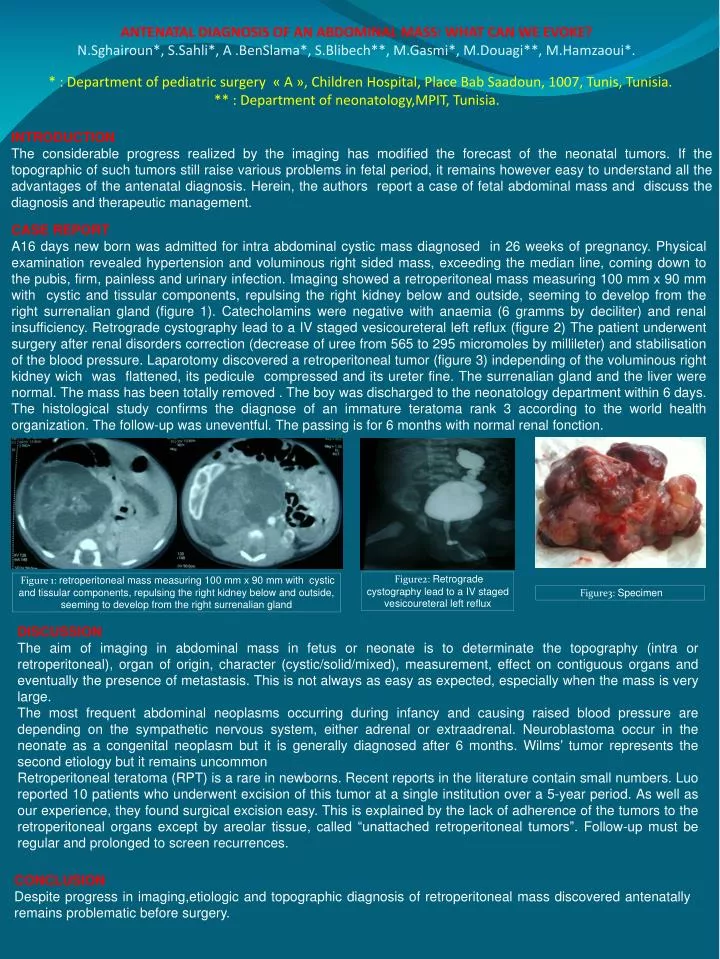
ANTENATAL DIAGNOSIS OF AN ABDOMINAL MASS: WHAT CAN WE EVOKE? N.Sghairoun*, S.Sahli*, A .BenSlama*, S.Blibech**, M.Gasmi*, M.Douagi**, M.Hamzaoui*. * : Department of pediatricsurgery « A », ChildrenHospital, Place BabSaadoun, 1007, Tunis, Tunisia. ** : Department of neonatology,MPIT, Tunisia. INTRODUCTION The considerableprogressrealized by the imaging has modified the forecast of the neonataltumors. If the topographicof suchtumorsstillraisevariousproblems in fetalperiod, itremainshowevereasy to understand all the advantages of the antenataldiagnosis. Herein, the authors report a case of fetal abdominal mass and discuss the diagnosis and therapeutic management. • CASE REPORT • A16 days new bornwasadmitted for intra abdominal cystic mass diagnosed in 26 weeks of pregnancy. Physicalexaminationrevealed hypertension and voluminous right sided mass, exceeding the median line, coming down to the pubis, firm, painless and urinary infection.Imaging showed a retroperitoneal mass measuring 100 mm x 90 mm withcystic and tissular components, repulsing the right kidneybelow and outside, seeming to developfrom the right surrenalian gland (figure 1). Catecholaminswerenegativewithanaemia (6 gramms by deciliter) and renalinsufficiency. Retrogradecystographylead to a IV stagedvesicoureteralleft reflux (figure 2) The patient underwentsurgeryafterrenaldisorders correction (decrease of ureefrom565 to 295 micromoles by millileter) and stabilisation of the blood pressure.Laparotomydiscovered a retroperitonealtumor (figure 3)independing of the voluminous right kidneywichwasflattened, itspediculecompressedand itsureter fine. The surrenalian gland and the liverwere normal. The mass has been totallyremoved . The boy wasdischarged to the neonatologydepartmentwithin 6 days. The histologicalstudyconfirms the diagnose of an immature teratomarank 3 according to the world healthorganization.The follow-up was uneventful. The passing is for 6 months with normal renal fonction. Figure2: Retrogradecystographylead to a IV stagedvesicoureteralleft reflux Figure 1: retroperitoneal mass measuring 100 mm x 90 mm withcystic and tissular components, repulsing the right kidneybelow and outside, seeming to developfrom the right surrenalian gland Figure3: Specimen DISCUSSION The aim of imaging in abdominal mass in fetus or neonate is to determinate the topography (intra or retroperitoneal), organ of origin, character (cystic/solid/mixed), measurement, effect on contiguousorgans and eventually the presence of metastasis. This is not always as easy as expected, especially when the mass is very large. The most frequent abdominal neoplasms occurring during infancy and causing raised blood pressure are depending on the sympathetic nervous system, either adrenal or extraadrenal. Neuroblastoma occur in the neonate as a congenital neoplasm but it is generally diagnosed after 6 months. Wilms’ tumor represents the second etiology but it remains uncommon Retroperitoneal teratoma (RPT) is a rare in newborns. Recent reports in the literature contain small numbers. Luo reported 10 patients who underwent excision of this tumor at a single institution over a 5-year period. As well as our experience, they found surgical excision easy. This is explained by the lack of adherence of the tumors to the retroperitoneal organs except by areolar tissue, called “unattached retroperitoneal tumors”. Follow-up must beregular and prolonged to screenrecurrences. CONCLUSION Despiteprogress in imaging,etiologic and topographicdiagnosis of retroperitoneal mass discoveredantenatallyremainsproblematicbeforesurgery.