DERMATOPATHOLOGY (FOR DENTISTRY STUDENT)
530 likes | 951 Vues
DERMATOPATHOLOGY (FOR DENTISTRY STUDENT). Prateep Warnnissorn Department of Medicine, Naresuan University. *เพื่อเลี่ยงปัญหาการละเมิดลิขสิทธิ์จากการนำเอกสารนี้ขึ้นเวป จึงได้ทำลิงก์รูป. Overview Definition of terms Infectious diseases -wart, molluscum contagiousum

DERMATOPATHOLOGY (FOR DENTISTRY STUDENT)
E N D
Presentation Transcript
DERMATOPATHOLOGY(FOR DENTISTRY STUDENT) • Prateep Warnnissorn • Department of Medicine, Naresuan University *เพื่อเลี่ยงปัญหาการละเมิดลิขสิทธิ์จากการนำเอกสารนี้ขึ้นเวป จึงได้ทำลิงก์รูป
Overview • Definition of terms • Infectious diseases -wart, molluscum contagiousum • Inflammatory diseases -acne, urticaria, eczema, arthropod bites, psoriasis, seborrheic dermatitis, vesiculobullous disease • Neoplastic diseases -SCC, BCC & MM • Principles of therapy of skin change and pathology
OVERVIEW • Normal skin • Wound healing
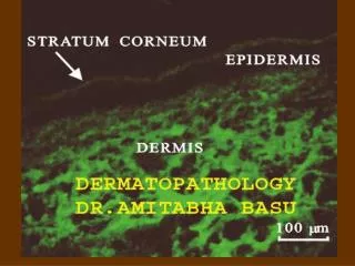
NORMAL SKIN • Epidermis • Dermis • Subcutaneous • Adnexal structures
EPIDERMIS http://headandneckcancerguide.org/wp-content/uploads/2013/02/61_epidermis2.jpg http://missinglink.ucsf.edu/lm/dermatologyglossary/img/Dermatology%20Glossary/Glossary%20Histo%20Images/Epidermis_40x-211.jpg
EPIDERMIS -MUCOUS MEMBRANE • S.spinosum • larger cells • pale cytoplasm-more glycogen • Granular layer -none • Keratinization with parakeratosis (retain nucleus) http://www.lab.anhb.uwa.edu.au/mb140/corepages/oral/oral.htm
DERMIS http://kreativestudios.com/Tooltip/05Integument/images/03Dermis.jpg
DERMIS http://www.lab.anhb.uwa.edu.au/mb140/corepages/connective/Images/skk040evg.jpg http://www.google.com/imgres?client=safari&rls=en&biw=1135&bih=669&tbm=isch&tbnid=n1dYl8VhB3YE1M%3A&imgrefurl=http%3A%2F%2Fwww.studyblue.com%2Fnotes%2Fnote%2Fn%2Funit-4-histology--connective-tissue-proper%2Fdeck%2F1989056&docid=5yd0dxzL1qRYPM&imgurl=http%3A%2F%2Fclassconnection.s3.amazonaws.com%2F844%2Fflashcards%2F1132844%2Fjpg%2Fcardimage_1989056_01327949756872.jpg&w=1000&h=688&ei=ab4nU4DdO4WFrAfghYCgDA&zoom=1&ved=0COABEIQcMC0&iact=rc&dur=509&page=3&start=30&ndsp=18
HYPODERMIS & ADNEXAL STRUCTURES http://apbrwww5.apsu.edu/thompsonj/Anatomy%20&%20Physiology/2010/2010%20Exam%20Reviews/Exam%202%20Review/dermis_diagram.gif
WOUND HEALING • Coagulation • Inflammatory • Proliferative & migration • Remodeling http://img.medscape.com/article/782/469/782469-fig2.jpg
DEFINITION OF TERMS • S.corneum/keratin -orthokeratosis, parakeratosis • S.malphigii -atrophy, acanthosis, papillomatosis • S.granular -hypergranulosis, hypogranulosis • S.spinusum -ballooning, spongiosis, acantholysis, dyskeratosis • Terms in viral infections • Exocytosis -white blood cells in epidermis
ORTHOKERATOSIS • Thickening of the s.corneum http://escholarship.org/uc/item/1qg46424/111902-3b.jpg
PARAKERATOSIS • Flattened keratinocyte nuclei in S.corneum http://www.skinpathology.org/psoriasis/
ATROPHY • Thining of epidermis http://www.globalskinatlas.com/upload/lg259_8.jpg
ACANTHOSIS • hypertrophy of s.spinosum http://classconnection.s3.amazonaws.com/866/flashcards/880334/png/acanthosis.png
PAPILLOMATOSIS • nipplelike growths http://www.ijdvl.com/articles/2013/79/3/images/ijdvl_2013_79_3_338_110769_f6.jpg
HYPERGRANULOSIS • thickened granular layer http://www.dermaamin.com/site/images/histo-pic/h/hypertrophic-lichen-planus/hypertrophic-lichen-planus1.jpg
HYPOGRANULOSIS • decrease thickness of granular layer http://openi.nlm.nih.gov/detailedresult.php?img=3276782_ad-23-S303-g002&req=4
INTRACELLULAR EDEMA • ballooning • intracellular clear round spaces • pale, large cells https://web.duke.edu/pathology/Week19/Week19.html#answer
INTERCELLULAR EDEMA • spongiosis • widening of intercellular bridge http://1.bp.blogspot.com/_51yzVOvZWEc/SzxAJJVLbiI/AAAAAAAAFjg/0PpWqE3_Pq0/s400/Eczema%20nummular2.jpg
ACANTHOLYSIS • separation and rounding up of cells • loss of intercellular adhesions http://missinglink.ucsf.edu/lm/dermatologyglossary/img/Dermatology%20Glossary/Glossary%20Histo%20Images/acantholysis.jpg
DYSKERATOSIS • necrosis, apoptosis • prematurely keratinised • prominent eosinophilic cytoplasm
TERMS IN VIRAL INFECTIONS • Coarse clumping keratohyaline granules • Intranuclear inclusion body • Intracytoplasmic inclusion body • multinucleated giant cells • Koilocyte
COARSE CLUMPING KERATOHYALINE GRANULES • large keratohyaline granules in s.granular layer http://forum.mckeedermpath.com/uploads/88ca6edbc0492119d1735034a98b55a8.jpg
INTRANUCLEAR INCLUSION BODY • large intranuclear inclusion body • eccentric, crescent bluish nucleus http://upload.wikimedia.org/wikipedia/commons/d/d3/Molluscum_contagiosum_1.jpg
INTRACYTOPLASMIC INCLUSION BODY http://cai.md.chula.ac.th/chulapatho/chulapatho/lecturenote/infection/Pathology%20of%20infection/inclusion%20bodies/verruca_vulgaris/verruca4.gif
MULTINUCLEATED GIANT CELL http://cai.md.chula.ac.th/chulapatho/chulapatho/lecturenote/infection/Pathology%20of%20infection/inclusion%20bodies/herpesimplex_files/SKIN085.jpg
VACUOLATED CELL /KOILOCYTE • perinuclear halo or clear cytoplasm http://jcwl.glmc.edu.cn/glblnet/severbj1/bl/JPEG3/SKIN036.JPG
INFECTIOUS DISEASES • Warts • Molluscum contagiosum
WARTS / VERRUCA VULGARIS • acanthosis • “church spire papillomatosis • ortho-and parakeratosis • hypergranulosis • koilocytosis • inward bending of tete http://cai.md.chula.ac.th/chulapatho/chulapatho/lecturenote/infection/Pathology%20of%20infection/inclusion%20bodies/verrusca_vulgaris.html
MOLLUSCUM CONTAGIOSUM • intracytoplasmic inclusion body • crescentric nucleus http://www.webpathology.com/slides/slides/ExtGenitalia_Molluscum1.jpg
INFLAMMATORY DISEASES • acne • urticaria • eczema • arthropod bites • psoriasis • seborrheic dermatitis • vesiculobullous disease -pemphigus, pemphigoid
ACNE VULGARIS http://accessmedicine.mhmedical.com/content.aspx?bookid=392§ionid=41138785 http://dermaamin.com/site/images/histo-pic/a/acne-vulgaris/acne-vulgaris1.jpg
URTICARIA • dermal edema • vascular dilatation & endothelial swelling • scant perivascular infiltration http://www.dermpedia.org/files/images/Urticaria_4.jpg
ECZEMA • spongiosis (acute > subacute) • spongiotic vesicle (acute > subacute) • exocytosis (acute > subacute) • parakeratosis (subacute) http://upload.wikimedia.org/wikipedia/commons/9/9d/Spongiotic_dermatitis_(2)_Dyshidrotic_.JPG
ECZEMA (CHRONIC) • vertical collagen streak • compact orthokeratosis • acanthosis (chronic > subacute) http://missinglink.ucsf.edu/lm/dermatologyglossary/img/Dermatology%20Glossary/Glossary%20Histo%20Images/lichen_simplex_chronicus_from_normal_skin_mid_power.jpg http://missinglink.ucsf.edu/lm/dermatologyglossary/img/Dermatology%20Glossary/Glossary%20Histo%20Images/lichen_simplex_chronicus_from_normal_skin_mid_power.jpg
ARTHROPOD BITE • edema epidermis & dermis • wedge shape inflammation • infiltration superficial & deep • mainly eosinophils http://classconnection.s3.amazonaws.com/352/flashcards/717352/png/untitled1319594530915.png
http://www.ijdvl.com/articles/2013/79/3/images/ijdvl_2013_79_3_338_110769_f4.jpghttp://www.ijdvl.com/articles/2013/79/3/images/ijdvl_2013_79_3_338_110769_f4.jpg PSORIASIS • parakeratosis & orthokeratosis • neutrophils • in s.corneum (Munro microabscess) • in upper epidermis (spongioform pustule of Kogoj) • thin suprapapillary plate (Auspitz sign) • dilate vessels close to the epidermis • elongation of rete ridge (camel foot) http://www.dermweb.com/pathology/graphics/annotations/psoriasis1ann.jpg http://www.ijdvl.com/articles/2013/79/3/images/ijdvl_2013_79_3_338_110769_f4.jpg
SEBORRHEIC DERMATITIS • eczema + psoriasis • spongiosis without spongiotic vesicle • shoulder parakeratosis (follicular ostia), neutrophilic parakeratosis • sebaceous gland -large & numerous http://dermaamin.com/site/images/histo-pic/s/seborrheic-dermatitis/seborrheic-dermatitis4.jpg
PEMPHIGUS VULGARIS • suprabasal separation “raw of tomb stone” • Direct immunoflourescence -IgG on surface of keratinocytes http://dermaamin.com/site/images/histo-pic/p/pemphigus-histopathology/pemphigus-histopathology25.jpg http://www.google.com/url?sa=i&source=images&cd=&docid=durhjvZDMNVQ8M&tbnid=7F2_20A-gcmTcM:&ved=0CAUQjBw4Og&url=http%3A%2F%2Fimg.medscape.com%2Fpi%2Femed%2Fckb%2Fdermatology%2F1048885-1064187-3368.jpg&ei=qSctU4XFOMbXrQfVp4CAAg&psig=AFQjCNF5H0fq_2qRG6pU9RqeDU9pd-Qx7w&ust=1395554602033805 http://www.ijdvl.com/articles/2013/79/3/images/ijdvl_2013_79_3_338_110769_f27.jpg
BULLOUS PEMPHIGOID • subepidermal separation • eosinophil • DIF -linear C3 & IgG at basement membrane zone http://plaza.umin.ac.jp/pathol2/photo-library/system/data/image_data/11348999517711.jpg http://eso-cdn.bestpractice.bmj.com/best-practice/images/bp/en-gb/453-8_default.jpg
NEOPLASM • Basal cell carcinoma • Squamous cell carcinoma • Malignant melanoma
BASAL CELL CARCINOMA • large uniform nuclei and scant cytoplasm • retraction of the stroma around the tumor islands -clefts • aggregations of basaloid keratinocytes extend from epidermis to dermis • peripheral palisading http://plaza.umin.ac.jp/pathol2/photo-library/system/data/image_data/11349004874921.jpg
SQUAMOUS CELL CARCINOMA • normal & atypical squamous cells -nuclear pleomorphism, apoptosis, mitosis • horn pearls -foci of keratinization in concentric rings of squamous cells • extension of atypical keratinocyte beyond the basement membrane (invasive SCC) http://missinglink.ucsf.edu/lm/dermatologyglossary/img/Dermatology%20Glossary/Glossary%20Histo%20Images/squamous_cell_carcinoma_invasive_high_power.jpg
MALIGNANT MELANOMA • architectural • asymmetry • poor lateral circumscription (single, scattered cells) • large size >5 mm) • epidermal nest larger than dermal nest • pagetoid spreading -spread to upper epidermis and later to dermis http://melanocytepathology.com/uploads/editor/Malignant_melanoma_12_a.jpg
MM • cytologic atypia • large nuclear & cytoplasm (with dusty pigment) • nuclear pleomorphism, hyperchromasia & variability • mitosis & necrotic https://secure.health.utas.edu.au/intranet/cds/pathprac/Files/Cases/Skin/Case96/Case96.htm
PRINCIPLES OF THERAPY OF SKIN CHANGE AND PATHOLOGY • Corticosteroids • Retinoids • Immunomodulators • Keratolytics • Antiperspiration • Psoriasis therapies
CORTICOSTEROIDS • Anti-proliferative • inhibit DNA synthesis • reduce keratinocyte size and proliferation • Anti-inflammatory • inhibit phospholiapse A2 release & transcription factors -activator protein-1 & nuclear factor kappa B • Immunosuppressive • suppress leukocyte migration, mast cells, Langerhans cells & T-cell proliferation • Vasoconstrictive
RETINOIDS • Increase • differential markers -involucrin, loricrin, filaggrin & transglutaminase • proliferation of basal keratinocytes
IMMUNOMODULATORS • Calcineurin inhibitor • tacrolimus • interact FK-506 binding protein, inhibit nuclear translocation • anti-inflammatory -decrease T-cell proliferation and pro inflammatory cytokines • Imidazoquinoline amine • imiquimod -TLR-7 & TLR-8 agonist • enhances immune response -proinflammatory cytokines • antiviral (wart, molluscum), antitumoral (actinic keratosis, basal cell carcinoma)