Download

1 / 22
220 likes | 403 Vues
Phage Display Selected T59 Peptide Binding Characterization to PPyCl. Archit B. Sanghvi The University of Texas at Austin Department of Biomedical Engineering. synthetic material. Motivation: Promoting Bioactivity. Soft Tissue Engineering. Drug Delivery. Bioreactor Systems.

E N D
Phage Display Selected T59 Peptide Binding Characterization to PPyCl Archit B. Sanghvi The University of Texas at Austin Department of Biomedical Engineering
synthetic material Motivation: Promoting Bioactivity Soft Tissue Engineering Drug Delivery Bioreactor Systems
De novo synthesis Covalent modification Physical adsorption • polylactic acid-polylysine • QuirkR. (2001) Biomaterials , • 22: 865-872. • protein self-assembly • Zhang S.(1999) Reac. & Func. • Polym., 41:91-102. • amine functional groups • Xiao S.J. (2000) Langmuir., 14(19): • 5507-5516. + + + Copolymerization • Limitations: • complex chemical processing • alteration of bulk polymer properties • Limitations: • weak hydrophobic • interactions • non-specific binding • Limitations: • complex chemistries • need existing functional groups Current State-of-the-Art
Advantages: • Control orientation • No changes to bulk property • High affinity binding • Ease of application • Applied to various biomaterials • including existing FDA approved polymers Polymer-Specific Surface Modification Peptide isolation using phage display: Yellow peptide motifs Red cell adhesion molecules Green growth factors
A 10 mm 10 mm B left right: T59 peptide on PPyCl, PPyPSS, and PS T59 peptide binds to PPyCl and not to PPyPSS or to polystyrene (PS). T59 Phage/Peptide Binding to PPyCl- left right: T59 phage on PPyCl, random phage on PPyCl
Functional Cell Applications T59-GRGDS on PPyCl promotes PC12 cell adhesion. Negative control --serum-free condition T59-GRGDS -- serum-free condition 10 mm
Characterization of T59 Peptide Binding to PPyCl • Mechanism of binding • Role of specific amino acids • Mechanical/adhesive strength • Compliance properties (unbinding force) • Binding affinity (Ka) • Quantify stability
His Arg Asp Surface Coverage Analysis: Fluorescamine Assay • Reacts with freea-amino groups (NH2-C-COOH) • Evaluate bound T59 peptide (variants) using std. curve. analysis Fluorescamine Assay
Identifying Critical Amino Acids of T59 Peptide in Binding PPyCl BT59 GGGGGG-LDYFVI RBT59 GGGGGG-IVFYDL T59D THRTSTLGYFVI T59HR TGGTSTLDYFVI T59 THRTSTLDYFVI RT59 IVFYDLTSTHRT FT59 THRTST-GGGGGG n = 6
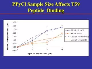
Evaluating the Role of Asp (8) in T59 Peptide Binding to PPyCl Input Conc. = 15 mM n = 6
Varying pH Effects T59 Peptide Binding to PPyCl n = 6 pKa of Asp R - group = 3.86
Characterization of T59 Peptide Binding to PPyCl • Mechanism of binding • Role of specific amino acids • Mechanical/adhesive strength • Compliance properties (unbinding force) • Binding affinity (Ka) • Quantify stability
Why are Adhesive Interactions Important? • Critical for cell function • Survival • Migration • Proliferation • Differentiation http://www.neuro.wustl.edu/neuromuscular/lab/adhesion.htm
What is an AFM? • Measures topography with a force probe (x,y,z dimensions) • Lateral resolution 0.5 nm • Vertical resolution 0.1 nm • Operates by measuring attractive or repulsive forces • Components • Cantilever-mounted tip • Piezoelectric micropositioner • Cantilever deflection sensor • Feedback micropositioner http://stm2.nrl.navy.mil/how-afm/how-afm.html#General%20concept http://www.asylumresearch.com/Products/Mfp3D/Mfp3D.shtml
Force Curve Analysis:Contact Mode AFM • Basic principle • Sharp probe (tip) raster-scans over the sample • Deflections are detected by optical methods • Air, high vacuum, and liquid • Control of forces used to study mechanical properties • Tip modification • Immobilization of receptor (T59 peptide) http://www.molec.com/index_what_is_afm.html Biotin-BSA Streptavidin Biotin-T59 PPyCl
Anatomy of a Force Curve Praster, C.B. et al. Probing nano-scale forces with the atomic force microscope. 1995, App. Notes. • Measures deflections off of the free end of the cantilever • FN = kNΔz • High force sensitivity (10-5 nN) • High dynamic range (0.0001-5000 nN)
-500 -600 pN -700 -800 -900 50 0 -50 -100 -150 -200 -250 nm 600 400 200 pN 0 -200 -400 -600 -200 -400 -600 -800 nm AFM Force Plots:T59-PPyCl & Streptavidin-Biotin surface binding approach T59 functionalized AFM tip approach to PPyCl surface ~128 pN retraction unbinding force Biotin functionalized AFM tip approach to streptavidin surface ~309 pN
Comparison of Adhesive Forces n = 10 spots/sample and 30 readings/spot Published Data biotin-streptavidin = 340 pN ± 120 Lee, G. et al. Langmuir 1994, 10. covalent bond (Au-S) = 1400 pN ± 300 Grandbois, M. et al. Science 1999, 283. oligonucleotide base pair (20) = 1520 pN Lee, G. et al. Science 1994, 266.
Conclusion • PPyCl-specific peptide sequence identified (“T59”) • Qualitative/quantitative demonstration of T59 phage/peptide binding • Functional application: T59-GRGDS modified PPyCl • Characterization of T59 peptide binding to PPyCl • Involvement of Asp (8) in binding PPyCl • Strength of binding (adhesion force)
synthetic material Motivation: Promoting Bioactivity Soft Tissue Engineering Drug Delivery Bioreactor Systems
Acknowledgements • Dr. Wolfgang Frey (BME, UT @ Austin) • John Mendenhall and Dr. Klaus Linse-Institute for Cellular and Molecular Biology (ICMB) • Dr. Yang-Ming Sun - Texas Materials Institute (TMI) • John Wright - IGERT • Funding - National Science Foundation and Gillson Longenbaugh Foundation