Download
1 / 24
260 likes | 325 Vues
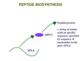
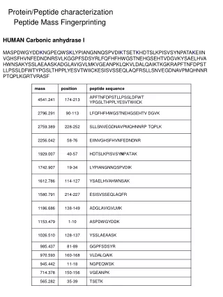
Protein/Peptide characterization. Peptide Mass Fingerprinting. HUMAN Carbonic anhydrase I.

E N D
Protein/Peptide characterization Peptide Mass Fingerprinting HUMAN Carbonic anhydrase I MASPDWGYDDKNGPEQWSKLYPIANGNNQSPVDIKTSETKHDTSLKPISVSYNPATAKEIINVGHSFHVNFEDNDNRSVLKGGPFSDSYRLFQFHFHWGSTNEHGSEHTVDGVKYSAELHVAHWNSAKYSSLAEAASKADGLAVIGVLMKVGEANPKLQKVLDALQAIKTKGKRAPFTNFDPSTLLPSSLDFWTYPGSLTHPPLYESVTWIICKESISVSSEQLAQFRSLLSNVEGDNAVPMQHNNRPTQPLKGRTVRASF
Ion Sources Matrix-assisted Laser Desorption/Ionization (MALDI)
Mass Analyzers “MS-only” Time-of-Flight (Tof) Nitrogen Laser 337 nm + + + + + + + + + + + + + + d Field-free drift region Detector -20,000 V MALDI plate + 20,000 V
Mass Analyzers Reflectron Tof, orthogonal injection
Reflectron Tof – increase in resolution Delayed Extraction – increase in resolution
432.9226 8000 7000 433.2556 6000 5000 4000 433.5887 3000 2000 1000 0 432.4 432.6 432.8 433.0 433.2 433.4 433.6 433.8 434.0 434.2 434.4 434.6 434.8 432.9226 8000 Intensity, counts 0 400 500 600 700 800 900 1000 1100 1200 1300 m/z, amu Angiotensin: DRVYIHPFHL C62H89N17O14 Dm/z = 0.33 Dm = 1 Da, z=3 Dm/z = 0.33 Intensity Mass to Charge ratio (m/z) “Monoisotopic Peak” 12C621H8914N1716O14 X + 2 peak 12C6013C21H8914N1716O14 + 12C6113C11H8914N1615N116O14 + 12C621H8914N1616O1318O1 X + 1 peak 12C6113C11H8914N1716O14 + 12C621H8914N1615N116O14
8000 77% 7000 6000 5000 4000 3000 2000 1000 0 432.4 432.6 432.8 433.0 433.2 433.4 433.6 433.8 434.0 434.2 434.4 434.6 434.8 Electron = 0.000549 g/mol Proton = 1.007253 g/mol Neutron = 1.00864 g/mol DRVYIHPFHL C62H89N17O14 [M+3H]3+ = 432.9226 x 3 = 1298.768 Measurement mass error = ~50 ppm X + 1 peak 12C6113C11H8914N1716O14 + 12C621H8914N1615N116O14 + 12C621H882H114N1716O14 68% C62H89N17O14 } 75% 6.3% 0.9%
Protein/Peptide characterization Peptide Mass Fingerprinting HUMAN Carbonic anhydrase I MASPDWGYDDKNGPEQWSKLYPIANGNNQSPVDIKTSETKHDTSLKPISVSYNPATAKEIINVGHSFHVNFEDNDNRSVLKGGPFSDSYRLFQFHFHWGSTNEHGSEHTVDGVKYSAELHVAHWNSAKYSSLAEAASKADGLAVIGVLMKVGEANPKLQKVLDALQAIKTKGKRAPFTNFDPSTLLPSSLDFWTYPGSLTHPPLYESVTWIICKESISVSSEQLAQFRSLLSNVEGDNAVPMQHNNRPTQPLKGRTVRASF
1079.4690 1078.9731 1079.9691 Intensity, counts 1079.0 1080.0 1081.0 m/z, amu 539.9927 540.2492 719.6340 540.4936 1400 Intensity, counts 540.7446 1300 540.9927 1200 539.9927 1100 540.0 540.4 540.8 541.2 m/z, amu 1000 900 800 Intensity, counts 700 563.9729 600 500 684.9795 400 1079.4690 597.6693 300 670.5904 200 100 0 550 600 650 700 750 800 850 900 950 1000 1050 m/z, amu Electrospray Ionization (ESI) [M+3H]3+ [M+4H]4+ [M+2H]2+
Protein/Peptide characterization Collision-activated dissociation (CAD) H R1 H H O R1 O R1 O N H2 N + N N + N N + O R2 O R2 O R2 H H H “b” “y” R1 R1 + N N + O H H “a” b-type 116 272 371 534 647 784 881 1028 1165 1296 D R V Y I H P F H L y-type 1296 1181 1025 926 763 650 513 416 269 132 b5 b62+ a62+ y62+ y2 a82+ b4 a5 y4 y1 y52+ b6
Introduction to Mass Spectrometry Amino Acid Residue Masses
b 157 214 329 442 539 686 785 884 981 1155 R G D L P F V V P R 1155 999 942 827 714 617 470 371 272 175 y
MS Scan Similar to LIM and SH3 protein 1 hypothetical protein MGC2477 MS/MS #1 MS/MS #4 3 400 800 1200 1800 1600 5 m/z 1 2 4 300 600 900 1200 1500 200 400 600 800 1000 1200 200 400 600 800 1000 1200 m/z m/z skeletal muscle tropomyosin Acyl-CoA-binding protein ??? MS/MS #5 MS/MS #2 MS/MS #3 200 400 600 800 1000 1200 200 400 600 800 1000 1200 m/z m/z ~10 seconds MS scan MS/MS#2 MS/MS#5 MS/MS#1 MS/MS#4 MS/MS#3 m/z
+ - - + Mass Analyzers Quadrupole Mass Spectrometer lens Ion source lens quadrupole detector F0 = U – V cos wt U = fixed (DC) potential V cos wt = applied RF, amplitude V and frequency wt
Mass Analyzers Triple Quadrupole “Single quad” “Triple quad” lens lens lens Ion source lens quadrupole quadrupole quadrupole detector Q1 Q3 Q2 Why triple quadrupole? - MS/MS analysis of peptides lens quadrupole quadrupole quadrupole detector Q1 Q3 Q2 scan m/z values (single quad) Pass 1 m/z value Fragment, pass all fragment ions
Mass Analyzers Quadrupole Ion Trap
816.63 100 95 997 90 950 85 900 80 850 75 800 70 750 65 700 60 650 55 600 50 550 Intensity, counts Relative Abundance 45 500 40 450 35 400 432.30 350 30 783.95 300 25 778.23 701.98 250 20 200 15 593.28 150 743.58 10 857.95 1223.77 100 5 50 0 0 997 950 900 850 800 750 700 650 600 550 Intensity, counts 500 450 400 350 300 LTQ Ion trap Quadrupole Tof ITMS + c ESI Full ms 496.8928 445.2570 543.7190 451.2044 539.5885 +4 487.5576 +3 744.8367 +5 493.0928 601.6043 730.8341 591.2612 526.87 1166.32 887.06 620.07 453.33 1114.11 954.77 1024.82 420 440 460 480 500 520 540 560 580 600 620 640 660 680 700 720 740 760 780 800 820 840 860 880 400 500 600 700 800 900 1000 1100 1200 m/z, amu m/z 1166.32 100 1174.80 95 +3 90 496.8928 85 0.33 80 75 70 497.2254 65 +2? +3? 60 0.33 55 50 +3 1143.44 Relative Abundance 45 1167.21 0.87 40 +1 1175.88 487.5576 35 497.5597 30 487.8905 25 493.0928 250 20 200 1144.31 1168.22 15 488.2278 150 497.8955 10 1176.78 1173.91 100 1153.83 5 50 1165.69 1148.21 1156.90 1182.83 0 0 487.0 488.0 489.0 490.0 491.0 492.0 493.0 494.0 495.0 496.0 497.0 498.0 499.0 500.0 501.0 502.0 1140 1145 1150 1155 1160 1165 1170 1175 1180 m/z, amu m/z
Fourier Transform Ion Cyclotron Resonance Mass Spectrometer (FTMS)
8000 7000 6000 5000 4000 3000 2000 1000 0 432.4 432.6 432.8 433.0 433.2 433.4 433.6 433.8 434.0 434.2 434.4 434.6 434.8 635.7516 49 Intensity, counts 636.2614 0 636.0 637.0 m/z, amu Introduction to Mass Spectrometry Resolution m/Dm Dm = FWHM resolution = 11,000 resolution = 5,000,000 Dynamic Range 432.9226 8490 8000 7500 7000 6500 6000 5500 5000 Intensity, counts 4500 4000 3500 3000 2500 2000 1500 382.2313 463.7657 1000 648.8623 858.5471 500 513.2979 0 400 450 500 550 600 650 700 750 800 850 900 950 1000 m/z, amu