Development of the Tetrapod Limb
410 likes | 1.34k Vues
BIOL 370 – Developmental Biology Topic #16. Development of the Tetrapod Limb. Lange. Pattern formation – study of the tangible (statistically), orderly outcomes of self-organization and the common principles behind similar patterns in nature.

Development of the Tetrapod Limb
E N D
Presentation Transcript
BIOL 370 – Developmental Biology Topic #16 Development of the Tetrapod Limb Lange
Pattern formation – study of the tangible (statistically), orderly outcomes of self-organization and the common principles behind similar patterns in nature. In developmental biology, pattern formation refers to the generation of complex organizations of cell fates in space and time. Pattern formation is controlled by genes.
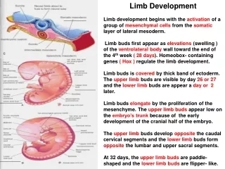
Periodicities of limb pattern formation: Stylopod humerus & femur Zeygopod ulna & radius / tibia & fibula Autopod carpals, metacarpals, & phalanges / tarsals, metatarsals, & phalanges
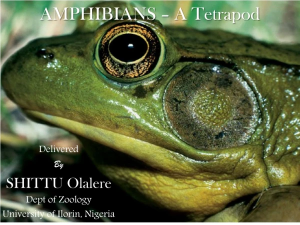
Figure 13.4Multilimbed Pacific tree frog (Hyla regilla), the result of infestation of the tadpole-stage developing limb buds by trematode cysts These cysts drive additional limb development when they feed on the limb bud leading to splitting of the bud into multiple growing regions.
Figure 13.5Fgf10 expression and action in the developing chick limb Fgf10 – fibroblast growth factor 10… proteins encoded by this gene are of the fibroblast growth factor (FGF) family. FGF family genes all possess broad based mitogenic and cell survival actions, including embryonic development.
Figure 13.6Molecular model for initiation of the limb bud in the chick between 48 and 54 hours of gestation Watch the process involving the Fgf10 gene.
Figure 13.7Specification of limb type in the chick by Tbx4 and Tbx5 (Part 1) Tbx4 – T-box 4 gene… hind limb development, Tbx5 – T-box 5 gene… forelimb development.
Figure 13.7Specification of limb type in the chick by Tbx4 and Tbx5 (Part 2) In the study in (B) we see a study in which an FGF bead is inserted in the area between the action of Tbx4 and Tbx5 resulting in an additional limb that is chimeric.
Figure 13.9Summary of experiments demonstrating the effect of the apical ectodermal ridge (AER) on the underlying mesenchyme Apical Ectodermal Ridge - a structure that forms from the ectodermal cells at the distal end of each limb bud and acts as a major signaling center to ensure proper development of a limb. After the limb bud induces AER formation, the AER and limb mesenchyme—including the zone of polarizing activity (ZPA)—continue to communicate with each other to direct further limb development
Figure 13.10Fgf8 in the apical ectodermal ridge Fgf8 – fibroblast growth factor 8… important within AER action
Figure 13.12Control of proximal-distal specification by the progress zone mesenchyme • Transplantation of an early wing bud progress zone to a late wing bud zone. • Transplant of a late wing bud progress zone to an early wing bud zone Extra radius & ulna No extra development
Figure 13.15When a ZPA is grafted to anterior limb bud mesoderm, duplicated digits emerge as a mirror image of the normal digits
Figure 13.16Sonic hedgehog protein is expressed in the ZPA ZPA - Zone of Polarizing Activity… an area of mesenchyme that releases signals instructing the developing limb bud to form along its anterior/posterior axis.
Figure 13.17Ectopic expression of mouse sonic hedgehog in the anterior limb causes extra digit formation The mutant form in (B) is called the Hx mutation (hemimelic extratoes). Shh expression that is ectopic (abnormal) can result in polydactyl digits in these mice.
Figure 13.18Deletion of limb bone elements by the deletion of paralogous Hox genes Notice how the differential expression of Hox prologues may shape overall development, but that there are still many similarities.
Figure 13.18Deletion of limb bone elements by the deletion of paralogous Hox genes (Part 4) Human Synpolydactyly - (the term means simply, “many fingers joined together”)… a result of homozygosity at the HoxD-13 loci. In people with this syndrome, there is also often malformations of the urogenital system, which also expresses this gene.
Ernest Hemingway’s home (now a museum) in Key West Florida houses descendants of his own pet polydactyl cats. In cat breeding circles, the polydactyl cats are sometimes called Hemmingway cats.
Figure 13.22Regulation of digit identity by BMP concentrations in the interdigital space anterior to the digit and by Gli3 Gli3 ( producing Zinc finger protein Gli3) represses dHand and Gremlin, which are involved in developing digits. This experiment shows a potential mechanism for “webbing” of digits.
Figure 13.26Patterns of cell death in leg primordia of (A) duck and (B) chick embryos Result is a webbed foot. Result is a non-webbed foot.
Figure 13.27Autopods of chicken feet and duck feet are shown at similar stages The expression of Gremlin in the duck (arrows) is thought to prevent the full apoptotic death of the webbing in the duck.
Figure 13.28Inhibition of cell death by inhibiting BMPs Gremlin protein soaked beads placed in the mesodermal webbing of (B) show persistence of the webbing.
Figure 13.29Possible involvement of BMPs in stabilizing cartilage and apoptosis BMP and noggin expression may be the cause of the cartilage defects showin in (C).
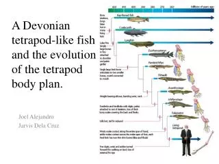
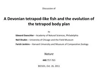
Figure 13.30Tiktaalik, a fish with wrists and fingers, lived in shallow waters ~375 million years ago Tiktaalik - a monospecific genus of extinct sarcopterygian (lobe-finned) fish from the late Devonian period, with many features akin to those of tetrapods. Originally these were first discovered in northern Canada on Ellesmere Island.